


Neutron structure of alginate lysase PsPL7C from Paradendryphiella salina soaked with penta-mannuronic acid
Meilleur, F., Morth, J.P., Wilkens, C.To be published.
Experimental Data Snapshot
wwPDB Validation 3D Report Full Report

Entity ID: 1 | |||||
|---|---|---|---|---|---|
| Molecule | Chains | Sequence Length | Organism | Details | Image |
| Alginate lyase | 234 | Paradendryphiella salina | Mutation(s): 0 Gene Names: PsAlg7C | 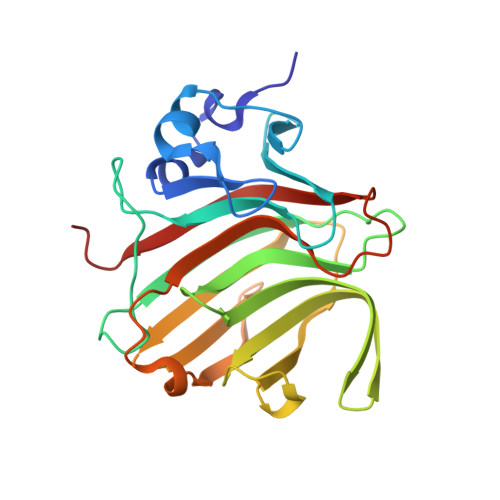 | |
UniProt | |||||
Find proteins for A0A7I9C8Z1 (Paradendryphiella salina) Explore A0A7I9C8Z1 Go to UniProtKB: A0A7I9C8Z1 | |||||
Entity Groups | |||||
| Sequence Clusters | 30% Identity50% Identity70% Identity90% Identity95% Identity100% Identity | ||||
| UniProt Group | A0A7I9C8Z1 | ||||
Sequence AnnotationsExpand | |||||
| |||||
Entity ID: 2 | |||||
|---|---|---|---|---|---|
| Molecule | Chains | Length | 2D Diagram | Glycosylation | 3D Interactions |
| 4-deoxy-alpha-L-erythro-hex-4-enopyranuronic acid-(1-4)-beta-D-mannopyranuronic acid | B | 2 |  | N/A | |
Entity ID: 3 | |||||
|---|---|---|---|---|---|
| Molecule | Chains | Length | 2D Diagram | Glycosylation | 3D Interactions |
| beta-D-mannopyranuronic acid-(1-4)-alpha-D-mannopyranuronic acid-(1-4)-beta-D-mannopyranuronic acid | C | 3 |  | N/A | |
| Length ( Å ) | Angle ( ˚ ) |
|---|---|
| a = 42.089 | α = 90 |
| b = 61.451 | β = 90 |
| c = 91.882 | γ = 90 |
| Funding Organization | Location | Grant Number |
|---|---|---|
| Not funded | -- |
RCSB PDB (citation) is hosted by
RCSB PDB is a member of the

