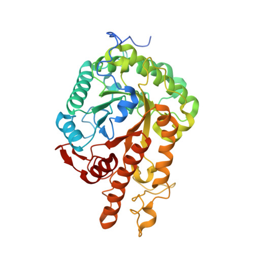
A Multiplexing Activity-Based Protein-Profiling Platform for Dissection of a Native Bacterial Xyloglucan-Degrading System.
McGregor, N.G.S., de Boer, C., Foucart, Q.P.O., Beenakker, T., Offen, W.A., Codee, J.D.C., Willems, L.I., Overkleeft, H.S., Davies, G.J.(2023) ACS Cent Sci 9: 2306-2314
- PubMed: 38161374
- DOI: https://doi.org/10.1021/acscentsci.3c00831
- Primary Citation of Related Structures:
8OZ1 - PubMed Abstract:
Bacteria and yeasts grow on biomass polysaccharides by expressing and excreting a complex array of glycoside hydrolase (GH) enzymes. Identification and annotation of such GH pools, which are valuable commodities for sustainable energy and chemistries, by conventional means (genomics, proteomics) are complicated, as primary sequence or secondary structure alignment with known active enzymes is not always predictive for new ones. Here we report a "low-tech", easy-to-use, and sensitive multiplexing activity-based protein-profiling platform to characterize the xyloglucan-degrading GH system excreted by the soil saprophyte, Cellvibrio japonicus , when grown on xyloglucan. A suite of activity-based probes bearing orthogonal fluorophores allows for the visualization of accessory exo -acting glycosidases, which are then identified using biotin-bearing probes. Substrate specificity of xyloglucanases is directly revealed by imbuing xyloglucan structural elements into bespoke activity-based probes. Our ABPP platform provides a highly useful tool to dissect xyloglucan-degrading systems from various sources and to rapidly select potentially useful ones. The observed specificity of the probes moreover bodes well for the study of other biomass polysaccharide-degrading systems, by modeling probe structures to those of desired substrates.
Organizational Affiliation:
Department of Chemistry, The University of York, Heslington, York YO10 5DD, United Kingdom.