Genetic and functional diversity of beta-N-acetylgalactosamine-targeting glycosidases expanded by deep-sea metagenome analysis.
Sumida, T., Hiraoka, S., Usui, K., Ishiwata, A., Sengoku, T., Stubbs, K.A., Tanaka, K., Deguchi, S., Fushinobu, S., Nunoura, T.(2024) Nat Commun 15: 3543-3543
- PubMed: 38730244
- DOI: https://doi.org/10.1038/s41467-024-47653-2
- Primary Citation of Related Structures:
8K2F, 8K2G, 8K2H, 8K2I, 8K2J, 8K2K, 8K2L, 8K2M, 8K2N - PubMed Abstract:
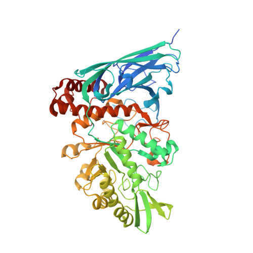
β-N-Acetylgalactosamine-containing glycans play essential roles in several biological processes, including cell adhesion, signal transduction, and immune responses. β-N-Acetylgalactosaminidases hydrolyze β-N-acetylgalactosamine linkages of various glycoconjugates. However, their biological significance remains ambiguous, primarily because only one type of enzyme, exo-β-N-acetylgalactosaminidases that specifically act on β-N-acetylgalactosamine residues, has been documented to date. In this study, we identify four groups distributed among all three domains of life and characterize eight β-N-acetylgalactosaminidases and β-N-acetylhexosaminidase through sequence-based screening of deep-sea metagenomes and subsequent searching of public protein databases. Despite low sequence similarity, the crystal structures of these enzymes demonstrate that all enzymes share a prototype structure and have diversified their substrate specificities (oligosaccharide-releasing, oligosaccharide/monosaccharide-releasing, and monosaccharide-releasing) through the accumulation of mutations and insertional amino acid sequences. The diverse β-N-acetylgalactosaminidases reported in this study could facilitate the comprehension of their structures and functions and present evolutionary pathways for expanding their substrate specificity.
Organizational Affiliation:
Research Center for Bioscience and Nanoscience, Japan Agency for Marine-Earth Science and Technology (JAMSTEC), Yokosuka, Japan. sumidat@jamstec.go.jp.