8JZN

Structure of a fungal 1,3-beta-glucan synthase


Experimental Data Snapshot

  • Method: ELECTRON MICROSCOPY
  • Resolution: 2.47 Å
  • Aggregation State: PARTICLE 
  • Reconstruction Method: SINGLE PARTICLE 

wwPDB Validation   3D Report Full Report


This is version 1.1 of the entry. See complete history


Literature

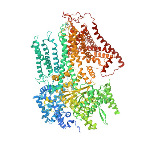
Structure of a fungal 1,3-beta-glucan synthase.

Zhao, C.R.You, Z.L.Chen, D.D.Hang, J.Wang, Z.B.Ji, M.Wang, L.X.Zhao, P.Qiao, J.Yun, C.H.Bai, L.

(2023) Sci Adv 9: eadh7820-eadh7820

  • DOI: https://doi.org/10.1126/sciadv.adh7820
  • Primary Citation of Related Structures:  
    8JZN

  • PubMed Abstract: 

    1,3-β-Glucan serves as the primary component of the fungal cell wall and is produced by 1,3-β-glucan synthase located in the plasma membrane. This synthase is a molecular target for antifungal drugs such as echinocandins and the triterpenoid ibrexafungerp. In this study, we present the cryo-electron microscopy structure of Saccharomyces cerevisiae 1,3-β-glucan synthase (Fks1) at 2.47-Å resolution. The structure reveals a central catalytic region adopting a cellulose synthase fold with a cytosolic conserved GT-A-type glycosyltransferase domain and a closed transmembrane channel responsible for glucan transportation. Two extracellular disulfide bonds are found to be crucial for Fks1 enzymatic activity. Through structural comparative analysis with cellulose synthases and structure-guided mutagenesis studies, we gain previously unknown insights into the molecular mechanisms of fungal 1,3-β-glucan synthase.


  • Organizational Affiliation

    Department of Biochemistry and Biophysics, School of Basic Medical Sciences, Peking University, Beijing, China.


Macromolecules
Find similar proteins by:  (by identity cutoff)  |  3D Structure
Entity ID: 1
MoleculeChains Sequence LengthOrganismDetailsImage
1,3-beta-glucan synthase component FKS11,876Saccharomyces cerevisiaeMutation(s): 0 
Gene Names: FKS1CND1CWH53ETG1GLS1GSC1PBR1YLR342WL8300.6
EC: 2.4.1.34
Membrane Entity: Yes 
UniProt
Find proteins for P38631 (Saccharomyces cerevisiae (strain ATCC 204508 / S288c))
Explore P38631 
Go to UniProtKB:  P38631
Entity Groups  
Sequence Clusters30% Identity50% Identity70% Identity90% Identity95% Identity100% Identity
UniProt GroupP38631
Sequence Annotations
Expand
  • Reference Sequence
Oligosaccharides

Help

Entity ID: 2
MoleculeChains Length2D Diagram Glycosylation3D Interactions
2-acetamido-2-deoxy-beta-D-glucopyranose-(1-4)-2-acetamido-2-deoxy-beta-D-glucopyranose
B
2N-Glycosylation
Glycosylation Resources
GlyTouCan:  G42666HT
GlyCosmos:  G42666HT
GlyGen:  G42666HT
Small Molecules
Ligands 3 Unique
IDChains Name / Formula / InChI Key2D Diagram3D Interactions
AV0
Query on AV0

Download Ideal Coordinates CCD File 
D [auth A],
E [auth A]
Lauryl Maltose Neopentyl Glycol
C47 H88 O22
MADJBYLAYPCCOO-VWHTXWAPSA-N
POV (Subject of Investigation/LOI)
Query on POV

Download Ideal Coordinates CCD File 
C [auth A],
T [auth A]
(2S)-3-(hexadecanoyloxy)-2-[(9Z)-octadec-9-enoyloxy]propyl 2-(trimethylammonio)ethyl phosphate
C42 H82 N O8 P
WTJKGGKOPKCXLL-PFDVCBLKSA-N
ERG
Query on ERG

Download Ideal Coordinates CCD File 
F [auth A]
G [auth A]
H [auth A]
I [auth A]
J [auth A]
F [auth A],
G [auth A],
H [auth A],
I [auth A],
J [auth A],
K [auth A],
L [auth A],
M [auth A],
N [auth A],
O [auth A],
P [auth A],
Q [auth A],
R [auth A],
S [auth A]
ERGOSTEROL
C28 H44 O
DNVPQKQSNYMLRS-APGDWVJJSA-N
Experimental Data & Validation

Experimental Data

  • Method: ELECTRON MICROSCOPY
  • Resolution: 2.47 Å
  • Aggregation State: PARTICLE 
  • Reconstruction Method: SINGLE PARTICLE 

Structure Validation

View Full Validation Report


Entry History & Funding Information

Deposition Data


Funding OrganizationLocationGrant Number
National Natural Science Foundation of China (NSFC)China32171212

Revision History  (Full details and data files)

  • Version 1.0: 2023-10-04
    Type: Initial release
  • Version 1.1: 2023-12-13
    Changes: Derived calculations, Structure summary