8JBO

Crystal structure of TxGH116 from Thermoanaerobacterium xylanolyticum with isofagomine


Experimental Data Snapshot

  • Method: X-RAY DIFFRACTION
  • Resolution: 2.00 Å
  • R-Value Free: 0.193 
  • R-Value Work: 0.155 
  • R-Value Observed: 0.157 

wwPDB Validation   3D Report Full Report


Ligand Structure Quality Assessment 


This is version 1.0 of the entry. See complete history


Literature

Structural basis for inhibition of a GH116 beta-glucosidase and its missense mutants by GBA2 inhibitors: Crystallographic and quantum chemical study.

Meelua, W.Thinkumrob, N.Saparpakorn, P.Pengthaisong, S.Hannongbua, S.Ketudat Cairns, J.R.Jitonnom, J.

(2023) Chem Biol Interact 384: 110717-110717

  • DOI: https://doi.org/10.1016/j.cbi.2023.110717
  • Primary Citation of Related Structures:  
    8JBO

  • PubMed Abstract: 

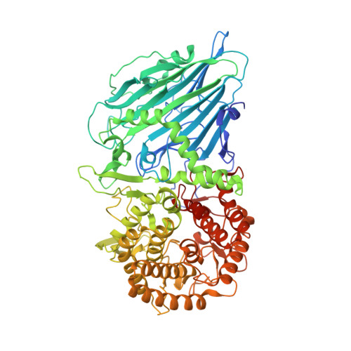
    The crystal structure of the Thermoanaerobacterium xylanolyticum in glycoside hydrolase family 116 (TxGH116) β-glucosidase provides a structural model for human GBA2 glucosylceramidase, an enzyme defective in hereditary spastic paraplegia and a potential therapeutic target for treating Gaucher disease. To assess the therapeutic potential of known inhibitors, the X-ray structure of TxGH116 in complex with isofagomine (IFG) was determined at 2.0 Å resolution and showed the IFG bound in a relaxed chair conformation. The binding of IFG and 7 other iminosugar inhibitors to wild-type and mutant enzymes (Asp508His and Arg786His) mimicking GBA2 pathogenic variants was then evaluated computationally by two-layered ONIOM calculations (at the B3LYP:PM7 level). Calculations showed that six charged residues, Glu441, Asp452, His507, Asp593, Glu777, and Arg786 influence inhibitor binding most. His507, Glu777 and Arg786, form strong hydrogen bonds with the inhibitors (∼1.4-1.6 Å). Thus, the missense mutation of one of these residues in Arg786His has a greater effect on the interaction energies for all inhibitors compared to Asp508His. In line with the experimental data for the inhibitors that have been tested, the favorable interaction energy between the inhibitors and the TxGH116 protein followed the trend: isofagomine > 1-deoxynojirimycin > glucoimidazole > N-butyl-deoxynojirimycin ≈ N-nonyl-deoxynojirimycin > conduritol B epoxide ≈ azepane 1 > azepane 2. The obtained structural and energetic properties and comparison to the GBA2 model can lead to understanding of structural requirement for inhibitor binding in GH116 to aid the design of high potency GBA2 inhibitors.


  • Organizational Affiliation

    Demonstration School, University of Phayao, Phayao, 56000, Thailand; Unit of Excellence in Computational Molecular Science and Catalysis, and Division of Chemistry, School of Science, University of Phayao, Phayao, 56000, Thailand.


Macromolecules
Find similar proteins by:  (by identity cutoff)  |  3D Structure
Entity ID: 1
MoleculeChains Sequence LengthOrganismDetailsImage
Glucosylceramidase
A, B
798Thermoanaerobacterium xylanolyticum LX-11Mutation(s): 0 
Gene Names: Thexy_2211
UniProt
Find proteins for F6BL85 (Thermoanaerobacterium xylanolyticum (strain ATCC 49914 / DSM 7097 / LX-11))
Explore F6BL85 
Go to UniProtKB:  F6BL85
Entity Groups  
Sequence Clusters30% Identity50% Identity70% Identity90% Identity95% Identity100% Identity
UniProt GroupF6BL85
Sequence Annotations
Expand
  • Reference Sequence
Small Molecules
Ligands 4 Unique
IDChains Name / Formula / InChI Key2D Diagram3D Interactions
IFM (Subject of Investigation/LOI)
Query on IFM

Download Ideal Coordinates CCD File 
C [auth A],
Q [auth B]
5-HYDROXYMETHYL-3,4-DIHYDROXYPIPERIDINE
C6 H13 N O3
QPYJXFZUIJOGNX-HSUXUTPPSA-N
GOL (Subject of Investigation/LOI)
Query on GOL

Download Ideal Coordinates CCD File 
AA [auth B]
E [auth A]
F [auth A]
G [auth A]
H [auth A]
AA [auth B],
E [auth A],
F [auth A],
G [auth A],
H [auth A],
I [auth A],
J [auth A],
K [auth A],
L [auth A],
M [auth A],
N [auth A],
R [auth B],
S [auth B],
T [auth B],
U [auth B],
V [auth B],
W [auth B],
X [auth B],
Y [auth B],
Z [auth B]
GLYCEROL
C3 H8 O3
PEDCQBHIVMGVHV-UHFFFAOYSA-N
EDO (Subject of Investigation/LOI)
Query on EDO

Download Ideal Coordinates CCD File 
BA [auth B],
O [auth A]
1,2-ETHANEDIOL
C2 H6 O2
LYCAIKOWRPUZTN-UHFFFAOYSA-N
CA (Subject of Investigation/LOI)
Query on CA

Download Ideal Coordinates CCD File 
CA [auth B],
D [auth A],
P [auth A]
CALCIUM ION
Ca
BHPQYMZQTOCNFJ-UHFFFAOYSA-N
Experimental Data & Validation

Experimental Data

  • Method: X-RAY DIFFRACTION
  • Resolution: 2.00 Å
  • R-Value Free: 0.193 
  • R-Value Work: 0.155 
  • R-Value Observed: 0.157 
  • Space Group: P 21 21 21
Unit Cell:
Length ( Å )Angle ( ˚ )
a = 54.791α = 90
b = 164.399β = 90
c = 179.614γ = 90
Software Package:
Software NamePurpose
REFMACrefinement
HKL-2000data collection
PDB_EXTRACTdata extraction
HKL-2000data reduction
HKL-2000data scaling
MOLREPphasing
Cootmodel building

Structure Validation

View Full Validation Report


Ligand Structure Quality Assessment 


Entry History & Funding Information

Deposition Data


Funding OrganizationLocationGrant Number
Other governmentThailandThailand Research Fund
Other governmentThailandSuranaree University of Technology
Other governmentThailandThailand Science Research and Innovation

Revision History  (Full details and data files)

  • Version 1.0: 2023-10-11
    Type: Initial release