8J52

Crystal structure of Flavihumibacter petaseus GH31 alpha-galactosidase mutant D304A in complex with alpha-1,4-galactobiose


Experimental Data Snapshot

  • Method: X-RAY DIFFRACTION
  • Resolution: 1.90 Å
  • R-Value Free: 0.192 
  • R-Value Work: 0.136 

wwPDB Validation   3D Report Full Report


Ligand Structure Quality Assessment 


This is version 1.1 of the entry. See complete history


Literature

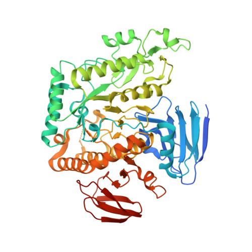
Structure-function analysis of bacterial GH31 alpha-galactosidases specific for alpha-(1→4)-galactobiose.

Ikegaya, M.Park, E.Y.Miyazaki, T.

(2023) FEBS J 290: 4984-4998

  • DOI: https://doi.org/10.1111/febs.16904
  • Primary Citation of Related Structures:  
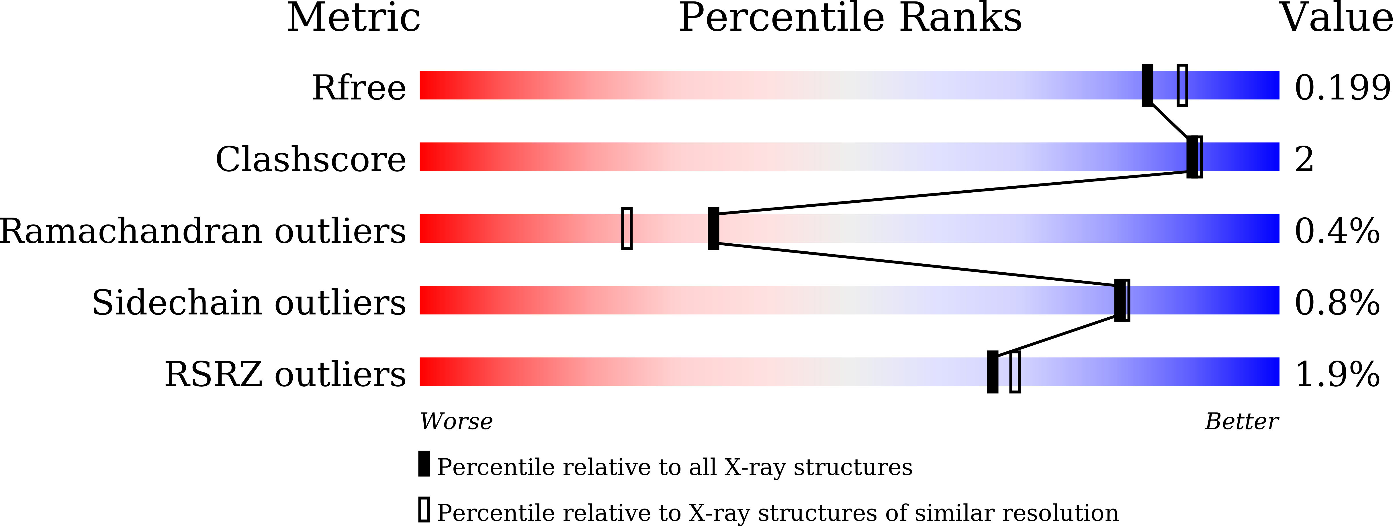
    8J50, 8J51, 8J52, 8J53

  • PubMed Abstract: 

    Glycoside hydrolase family 31 (GH31) contains α-glycoside hydrolases with different substrate specificities involved in various physiological functions. This family has recently been classified into 20 subfamilies using sequence similarity networks. An α-galactosidase from the gut bacterium Bacteroides salyersiae (BsGH31_19, which belongs to GH31 subfamily 19) was reported to have hydrolytic activity against the synthetic substrate p- nitrophenyl α-galactopyranoside, but its natural substrate remained unknown. BsGH31_19 shares low sequence identity (around 20%) with other reported GH31 α-galactosidases, PsGal31A from Pseudopedobacter saltans and human myogenesis-regulating glycosidase (MYORG), and was expected to have distinct specificity. Here, we characterized BsGH31_19 and its ortholog from a soil Bacteroidota bacterium, Flavihumibacter petaseus (FpGH31_19), and demonstrated that they showed high substrate specificity against α-(1→4)-linkages in α-(1→4)-galactobiose and globotriose [α-Gal-(1→4)-β-Gal-(1→4)-Glc], unlike PsGal31A and MYORG. The crystallographic analyses of BsGH31_19 and FpGH31_19 showed that their overall structures resemble those of MYORG and form a dimer with an interface different from that of PsGal31A and MYORG dimers. The structures of FpGH31_19 complexed with d-galactose and α-(1→4)-galactobiose revealed that amino acid residues that recognize a galactose residue at subsite +1 are not conserved between FpGH31_19 and BsGH31_19. The tryptophan (Trp153) that recognizes galactose at subsite -1 is homologous to the tryptophan residues in MYORG and α-galactosidases belonging to GH27, GH36, and GH97, but not in the bacterial GH31 member PsGal31A. Our results provide structural insights into molecular diversity and evolutionary relationships in the GH31 α-galactosidase subfamilies and the other α-galactosidase families.


  • Organizational Affiliation

    Department of Bioscience, Graduate School of Science and Technology, Shizuoka University, Japan.


Macromolecules
Find similar proteins by:  (by identity cutoff)  |  3D Structure
Entity ID: 1
MoleculeChains Sequence LengthOrganismDetailsImage
GH31 alpha-galactosidase
A, B
541Flavihumibacter petaseus NBRC 106054Mutation(s): 1 
Gene Names: FPE01S_01_01430
EC: 3.2.1.22
UniProt
Find proteins for A0A0E9MUN5 (Flavihumibacter petaseus NBRC 106054)
Explore A0A0E9MUN5 
Go to UniProtKB:  A0A0E9MUN5
Entity Groups  
Sequence Clusters30% Identity50% Identity70% Identity90% Identity95% Identity100% Identity
UniProt GroupA0A0E9MUN5
Sequence Annotations
Expand
  • Reference Sequence
Small Molecules
Ligands 4 Unique
IDChains Name / Formula / InChI Key2D Diagram3D Interactions
GAL (Subject of Investigation/LOI)
Query on GAL

Download Ideal Coordinates CCD File 
D [auth A],
L [auth B]
beta-D-galactopyranose
C6 H12 O6
WQZGKKKJIJFFOK-FPRJBGLDSA-N
GLA (Subject of Investigation/LOI)
Query on GLA

Download Ideal Coordinates CCD File 
C [auth A],
K [auth B]
alpha-D-galactopyranose
C6 H12 O6
WQZGKKKJIJFFOK-PHYPRBDBSA-N
EDO
Query on EDO

Download Ideal Coordinates CCD File 
E [auth A]
F [auth A]
G [auth A]
H [auth A]
I [auth A]
E [auth A],
F [auth A],
G [auth A],
H [auth A],
I [auth A],
M [auth B],
N [auth B],
O [auth B],
P [auth B],
Q [auth B],
R [auth B]
1,2-ETHANEDIOL
C2 H6 O2
LYCAIKOWRPUZTN-UHFFFAOYSA-N
CL
Query on CL

Download Ideal Coordinates CCD File 
J [auth A],
S [auth B]
CHLORIDE ION
Cl
VEXZGXHMUGYJMC-UHFFFAOYSA-M
Experimental Data & Validation

Experimental Data

  • Method: X-RAY DIFFRACTION
  • Resolution: 1.90 Å
  • R-Value Free: 0.192 
  • R-Value Work: 0.136 
  • Space Group: P 1
Unit Cell:
Length ( Å )Angle ( ˚ )
a = 71.499α = 101.922
b = 72.543β = 104.396
c = 81.202γ = 103.708
Software Package:
Software NamePurpose
REFMACrefinement
XDSdata reduction
Aimlessdata scaling
MOLREPphasing

Structure Validation

View Full Validation Report


Ligand Structure Quality Assessment 


Entry History & Funding Information

Deposition Data


Funding OrganizationLocationGrant Number
Japan Society for the Promotion of Science (JSPS)Japan19K15748

Revision History  (Full details and data files)

  • Version 1.0: 2023-07-26
    Type: Initial release
  • Version 1.1: 2023-11-01
    Changes: Data collection, Database references