8J3M

Structure of GH1 Br2 beta-glucosidase from bovine rumen metagenome


Experimental Data Snapshot

  • Method: X-RAY DIFFRACTION
  • Resolution: 2.00 Å
  • R-Value Free: 0.265 
  • R-Value Work: 0.233 

wwPDB Validation   3D Report Full Report


This is version 1.2 of the entry. See complete history

Re-refinement Note

A newer entry is available that reflects an alternative modeling of the original data: 8J5L


Literature

Structural and mutational analysis of glycoside hydrolase family 1 Br2 beta-glucosidase derived from bovine rumen metagenome.

Kaenying, W.Tagami, T.Suwan, E.Pitsanuwong, C.Chomngam, S.Okuyama, M.Kongsaeree, P.Kimura, A.Kongsaeree, P.T.

(2023) Heliyon 9: e21923-e21923

  • DOI: https://doi.org/10.1016/j.heliyon.2023.e21923
  • Primary Citation of Related Structures:  
    8J3M, 8J5L, 8J5M

  • PubMed Abstract: 

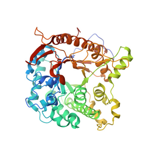
    Ruminant animals rely on the activities of β -glucosidases from residential microbes to convert feed fibers into glucose for further metabolic uses. In this report, we determined the structures of Br2, which is a glycoside hydrolase family 1 β -glucosidase from the bovine rumen metagenome. Br2 folds into a classical ( β / α ) 8 -TIM barrel domain but displays unique structural features at loop β 5→ α 5 and α -helix 5, resulting in different positive subsites from those of other GH1 enzymes. Br2 exhibited the highest specificity toward laminaritriose, suggesting its involvement in β -glucan hydrolysis in digested feed. We then substituted the residues at subsites +1 and + 2 of Br2 with those of Halothermothrix orenii β -glucosidase. The C170E and C221T mutations provided favorable interactions with glucooligosaccharide substrates at subsite +2, while the A219N mutation probably improved the substrate preference for cellobiose and gentiobiose relative to laminaribiose at subsite +1. The N407Y mutation increased the affinity toward cellooligosaccharides. These results give further insights into the molecular determinants responsible for substrate specificity in GH1 β -glucosidases and may provide a basis for future enzyme engineering applications.


  • Organizational Affiliation

    Department of Biochemistry, Faculty of Science, Kasetsart University, Bangkok 10900, Thailand.


Macromolecules
Find similar proteins by:  (by identity cutoff)  |  3D Structure
Entity ID: 1
MoleculeChains Sequence LengthOrganismDetailsImage
Beta-glucosidase
A, B, C, D
465uncultured bacteriumMutation(s): 0 
Gene Names: BG
UniProt
Find proteins for A0A1S5SJM8 (uncultured bacterium)
Explore A0A1S5SJM8 
Go to UniProtKB:  A0A1S5SJM8
Entity Groups  
Sequence Clusters30% Identity50% Identity70% Identity90% Identity95% Identity100% Identity
UniProt GroupA0A1S5SJM8
Sequence Annotations
Expand
  • Reference Sequence
Small Molecules
Ligands 2 Unique
IDChains Name / Formula / InChI Key2D Diagram3D Interactions
SO4
Query on SO4

Download Ideal Coordinates CCD File 
F [auth A]
G [auth A]
H [auth A]
J [auth B]
K [auth B]
F [auth A],
G [auth A],
H [auth A],
J [auth B],
K [auth B],
M [auth C],
N [auth C],
O [auth C],
P [auth C],
R [auth D]
SULFATE ION
O4 S
QAOWNCQODCNURD-UHFFFAOYSA-L
GOL
Query on GOL

Download Ideal Coordinates CCD File 
E [auth A],
I [auth B],
L [auth C],
Q [auth D]
GLYCEROL
C3 H8 O3
PEDCQBHIVMGVHV-UHFFFAOYSA-N
Experimental Data & Validation

Experimental Data

  • Method: X-RAY DIFFRACTION
  • Resolution: 2.00 Å
  • R-Value Free: 0.265 
  • R-Value Work: 0.233 
  • Space Group: P 21 21 21
Unit Cell:
Length ( Å )Angle ( ˚ )
a = 105.701α = 90
b = 113.873β = 90
c = 181.865γ = 90
Software Package:
Software NamePurpose
REFMACrefinement
XDSdata reduction
XDSdata scaling
PHENIXphasing

Structure Validation

View Full Validation Report


Entry History & Funding Information

Deposition Data


Funding OrganizationLocationGrant Number
Other governmentThailandNRCT5-RSA63002-09

Revision History  (Full details and data files)

  • Version 1.0: 2023-11-22
    Type: Initial release
  • Version 1.1: 2023-12-13
    Changes: Database references
  • Version 1.2: 2024-01-03
    Changes: Database references