8IYC

Structure insight into substrate recognition and catalysis by feruloyl esterase from Aspergillus sydowii


Experimental Data Snapshot

  • Method: X-RAY DIFFRACTION
  • Resolution: 1.55 Å
  • R-Value Free: 0.186 
  • R-Value Work: 0.134 
  • R-Value Observed: 0.137 

wwPDB Validation   3D Report Full Report


Ligand Structure Quality Assessment 


This is version 1.0 of the entry. See complete history


Literature

Structural insights into the molecular mechanisms of substrate recognition and hydrolysis by feruloyl esterase from Aspergillus sydowii.

Phienluphon, A.Kondo, K.Mikami, B.Nagata, T.Katahira, M.

(2023) Int J Biol Macromol 253: 127188-127188

  • DOI: https://doi.org/10.1016/j.ijbiomac.2023.127188
  • Primary Citation of Related Structures:  
    8IY8, 8IYB, 8IYC

  • PubMed Abstract: 

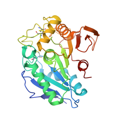
    The depolymerization of lignocellulosic biomass is facilitated by feruloyl esterases (FAEs), which hydrolyze ester bonds between lignin and polysaccharides. Fungal FAEs belonging to subfamily (SF) 6 release precursors such as ferulic acid derivatives, attractive for biochemical production. Among these, Aspergillus sydowii FAE (AsFaeE), an SF6 FAE, exhibits remarkable activity across various substrates. In this study, we conducted X-ray crystallography and kinetic analysis to unravel the molecular mechanisms governing substrate recognition and catalysis by AsFaeE. AsFaeE exhibits a typical α/β-hydrolase fold, characterized by a catalytic triad of serine, aspartate, and histidine. Comparative analysis of substrate-free, ferulic acid-bound, and sinapic acid-bound forms of AsFaeE suggests a conformational change in the loop covering the substrate-binding pocket upon binding. Notably, Pro158 and Phe159 within this loop cover the phenolic part of the substrate, forming three layers of planar rings. Our structure-based functional mutagenesis clarifies the roles of the residues involved in substrate binding and catalytic activity. Furthermore, distinct substrate-binding mechanisms between AsFaeE and other studied FAEs are identified. This investigation offers the initial structural insights into substrate recognition by SF6 FAEs, equipping us with structural knowledge that might facilitate the design of FAE variants capable of efficiently processing a wider range of substrate sizes.


  • Organizational Affiliation

    Institute of Advanced Energy, Kyoto University, Gokasho, Uji, Kyoto 611-0011, Japan; Graduate School of Energy Science, Kyoto University, Gokasho, Uji, Kyoto 611-0011, Japan.


Macromolecules
Find similar proteins by:  (by identity cutoff)  |  3D Structure
Entity ID: 1
MoleculeChains Sequence LengthOrganismDetailsImage
Feruloyl esterase
A, B
281Aspergillus sydowiiMutation(s): 0 
Gene Names: ASPSYDRAFT_1158585
UniProt
Find proteins for A0A1L9T9J3 (Aspergillus sydowii CBS 593.65)
Explore A0A1L9T9J3 
Go to UniProtKB:  A0A1L9T9J3
Entity Groups  
Sequence Clusters30% Identity50% Identity70% Identity90% Identity95% Identity100% Identity
UniProt GroupA0A1L9T9J3
Sequence Annotations
Expand
  • Reference Sequence
Small Molecules
Ligands 4 Unique
IDChains Name / Formula / InChI Key2D Diagram3D Interactions
SXX (Subject of Investigation/LOI)
Query on SXX

Download Ideal Coordinates CCD File 
C [auth A],
P [auth B]
SINAPINATE
C11 H12 O5
PCMORTLOPMLEFB-ONEGZZNKSA-N
NAG (Subject of Investigation/LOI)
Query on NAG

Download Ideal Coordinates CCD File 
D [auth A],
E [auth A],
Q [auth B],
R [auth B]
2-acetamido-2-deoxy-beta-D-glucopyranose
C8 H15 N O6
OVRNDRQMDRJTHS-FMDGEEDCSA-N
EDO (Subject of Investigation/LOI)
Query on EDO

Download Ideal Coordinates CCD File 
F [auth A]
G [auth A]
H [auth A]
I [auth A]
J [auth A]
F [auth A],
G [auth A],
H [auth A],
I [auth A],
J [auth A],
K [auth A],
L [auth A],
S [auth B],
T [auth B],
U [auth B],
V [auth B],
W [auth B],
X [auth B]
1,2-ETHANEDIOL
C2 H6 O2
LYCAIKOWRPUZTN-UHFFFAOYSA-N
ACT (Subject of Investigation/LOI)
Query on ACT

Download Ideal Coordinates CCD File 
AA [auth B]
BA [auth B]
CA [auth B]
M [auth A]
N [auth A]
AA [auth B],
BA [auth B],
CA [auth B],
M [auth A],
N [auth A],
O [auth A],
Y [auth B],
Z [auth B]
ACETATE ION
C2 H3 O2
QTBSBXVTEAMEQO-UHFFFAOYSA-M
Experimental Data & Validation

Experimental Data

  • Method: X-RAY DIFFRACTION
  • Resolution: 1.55 Å
  • R-Value Free: 0.186 
  • R-Value Work: 0.134 
  • R-Value Observed: 0.137 
  • Space Group: P 1 21 1
Unit Cell:
Length ( Å )Angle ( ˚ )
a = 37.378α = 90
b = 65.258β = 96.995
c = 95.381γ = 90
Software Package:
Software NamePurpose
BSSdata collection
XDSdata reduction
XDSdata scaling
MOLREPphasing
PHENIXrefinement

Structure Validation

View Full Validation Report


Ligand Structure Quality Assessment 


Entry History & Funding Information

Deposition Data


Funding OrganizationLocationGrant Number
Japan Society for the Promotion of Science (JSPS)Japan20H03192
Japan Society for the Promotion of Science (JSPS)Japan20K21477
Japan Society for the Promotion of Science (JSPS)Japan21H05519
Japan Society for the Promotion of Science (JSPS)Japan22H05596
Japan Society for the Promotion of Science (JSPS)Japan20K06524
Japan Society for the Promotion of Science (JSPS)Japan20K06164

Revision History  (Full details and data files)

  • Version 1.0: 2023-10-25
    Type: Initial release