


Crystal structure of UGT74AN3-UDP
Wei, H., Wei, H.To be published.
Experimental Data Snapshot
Entity ID: 1 | |||||
|---|---|---|---|---|---|
| Molecule | Chains | Sequence Length | Organism | Details | Image |
| Glycosyltransferase | 474 | Catharanthus roseus | Mutation(s): 0 EC: 2.4.1 | 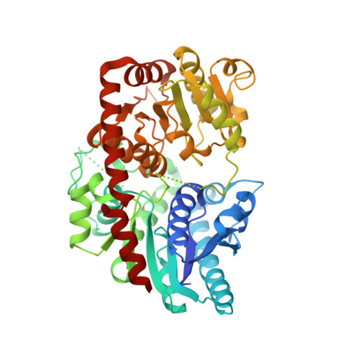 | |
UniProt | |||||
Find proteins for A0A385Z961 (Catharanthus roseus) Explore A0A385Z961 Go to UniProtKB: A0A385Z961 | |||||
Entity Groups | |||||
| Sequence Clusters | 30% Identity50% Identity70% Identity90% Identity95% Identity100% Identity | ||||
| UniProt Group | A0A385Z961 | ||||
Sequence AnnotationsExpand | |||||
| |||||
| Ligands 1 Unique | |||||
|---|---|---|---|---|---|
| ID | Chains | Name / Formula / InChI Key | 2D Diagram | 3D Interactions | |
| UPG (Subject of Investigation/LOI) Query on UPG | B [auth A] | URIDINE-5'-DIPHOSPHATE-GLUCOSE C15 H24 N2 O17 P2 HSCJRCZFDFQWRP-JZMIEXBBSA-N |  | ||
| Length ( Å ) | Angle ( ˚ ) |
|---|---|
| a = 45.437 | α = 90 |
| b = 52.068 | β = 90 |
| c = 200.195 | γ = 90 |
| Software Name | Purpose |
|---|---|
| PHENIX | refinement |
| Blu-Ice | data collection |
| XDS | data reduction |
| Aimless | data scaling |
| PHENIX | model building |
| PDB_EXTRACT | data extraction |
| Funding Organization | Location | Grant Number |
|---|---|---|
| National Natural Science Foundation of China (NSFC) | China | -- |
RCSB PDB (citation) is hosted by
RCSB PDB is a member of the

