The Distinctive Permutated Domain Structure of Periplasmic alpha-Amylase (MalS) from Glycoside Hydrolase Family 13 Subfamily 19.
An, Y., Tran, P.L., Yoo, M.J., Song, H.N., Park, K.H., Kim, T.J., Park, J.T., Woo, E.J.(2023) Molecules 28
- PubMed: 37241718
- DOI: https://doi.org/10.3390/molecules28103972
- Primary Citation of Related Structures:
8IM8 - PubMed Abstract:
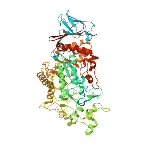
Periplasmic α-amylase MalS (EC. 3.2.1.1), which belongs to glycoside hydrolase (GH) family 13 subfamily 19, is an integral component of the maltose utilization pathway in Escherichia coli K12 and used among Ecnterobacteriaceae for the effective utilization of maltodextrin. We present the crystal structure of MalS from E. coli and reveal that it has unique structural features of circularly permutated domains and a possible CBM69. The conventional C-domain of amylase consists of amino acids 120-180 (N-terminal) and 646-676 (C-terminal) in MalS, and the whole domain architecture shows the complete circular permutation of C-A-B-A-C in domain order. Regarding substrate interaction, the enzyme has a 6-glucosyl unit pocket binding it to the non-reducing end of the cleavage site. Our study found that residues D385 and F367 play important roles in the preference of MalS for maltohexaose as an initial product. At the active site of MalS, β-CD binds more weakly than the linear substrate, possibly due to the positioning of A402. MalS has two Ca 2+ binding sites that contribute significantly to the thermostability of the enzyme. Intriguingly, the study found that MalS exhibits a high binding affinity for polysaccharides such as glycogen and amylopectin. The N domain, of which the electron density map was not observed, was predicted to be CBM69 by AlphaFold2 and might have a binding site for the polysaccharides. Structural analysis of MalS provides new insight into the structure-evolution relationship in GH13 subfamily 19 enzymes and a molecular basis for understanding the details of catalytic function and substrate binding of MalS.
Organizational Affiliation:
Disease Target Structure Research Center, Korea Research Institute of Bioscience and Biotechnology, Daejeon 34141, Republic of Korea.