


Crystal structure of alcohol oxidase PcAOX(M59V/Q60P/R61N)(Phanerochaete chrysosporium)
Wu, B., Wang, Y.To be published.
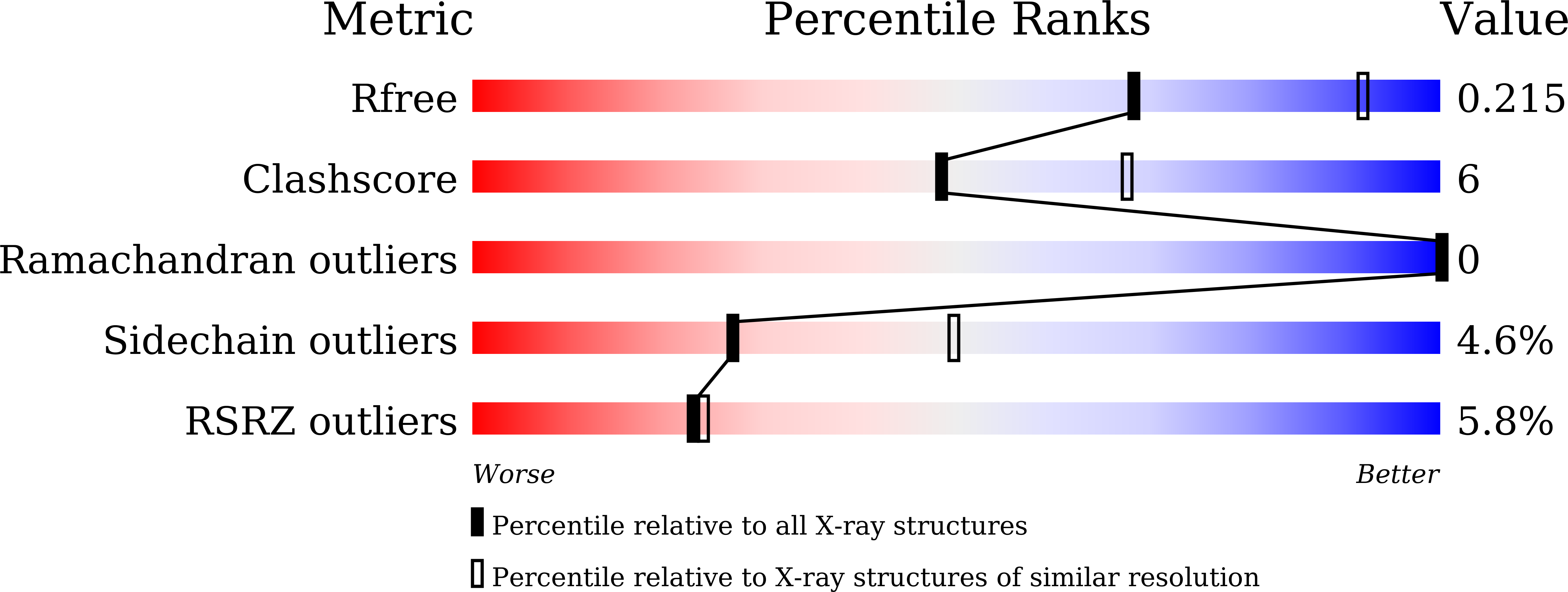
Experimental Data Snapshot
Entity ID: 1 | |||||
|---|---|---|---|---|---|
| Molecule | Chains | Sequence Length | Organism | Details | Image |
| Alcohol oxidase | 657 | Phanerodontia chrysosporium | Mutation(s): 3 Gene Names: AOX EC: 1.1.3.13 |  | |
UniProt | |||||
Find proteins for A0A977TIR6 (Phanerodontia chrysosporium) Explore A0A977TIR6 Go to UniProtKB: A0A977TIR6 | |||||
Entity Groups | |||||
| Sequence Clusters | 30% Identity50% Identity70% Identity90% Identity95% Identity100% Identity | ||||
| UniProt Group | A0A977TIR6 | ||||
Sequence AnnotationsExpand | |||||
| |||||
| Ligands 2 Unique | |||||
|---|---|---|---|---|---|
| ID | Chains | Name / Formula / InChI Key | 2D Diagram | 3D Interactions | |
| FAD (Subject of Investigation/LOI) Query on FAD | C [auth A], G [auth B] | FLAVIN-ADENINE DINUCLEOTIDE C27 H33 N9 O15 P2 VWWQXMAJTJZDQX-UYBVJOGSSA-N |  | ||
| SO4 (Subject of Investigation/LOI) Query on SO4 | D [auth A], E [auth A], F [auth A] | SULFATE ION O4 S QAOWNCQODCNURD-UHFFFAOYSA-L |  | ||
| Length ( Å ) | Angle ( ˚ ) |
|---|---|
| a = 161.26 | α = 90 |
| b = 161.26 | β = 90 |
| c = 113.34 | γ = 90 |
| Software Name | Purpose |
|---|---|
| PHENIX | refinement |
| autoPROC | data processing |
| HKL-2000 | data reduction |
| Coot | model building |
| PHASER | phasing |
| Funding Organization | Location | Grant Number |
|---|---|---|
| Not funded | -- |
RCSB PDB (citation) is hosted by
RCSB PDB is a member of the

