8IKX

An Arabidopsis polygalacturonase PGLR


Experimental Data Snapshot

  • Method: X-RAY DIFFRACTION
  • Resolution: 1.50 Å
  • R-Value Free: 0.179 
  • R-Value Work: 0.158 
  • R-Value Observed: 0.159 

wwPDB Validation   3D Report Full Report


Ligand Structure Quality Assessment 


This is version 1.1 of the entry. See complete history


Literature

A plant mechanism of hijacking pathogen virulence factors to trigger innate immunity.

Xiao, Y.Sun, G.Yu, Q.Gao, T.Zhu, Q.Wang, R.Huang, S.Han, Z.Cervone, F.Yin, H.Qi, T.Wang, Y.Chai, J.

(2024) Science 383: 732-739

  • DOI: https://doi.org/10.1126/science.adj9529
  • Primary Citation of Related Structures:  
    8IKW, 8IKX

  • PubMed Abstract: 

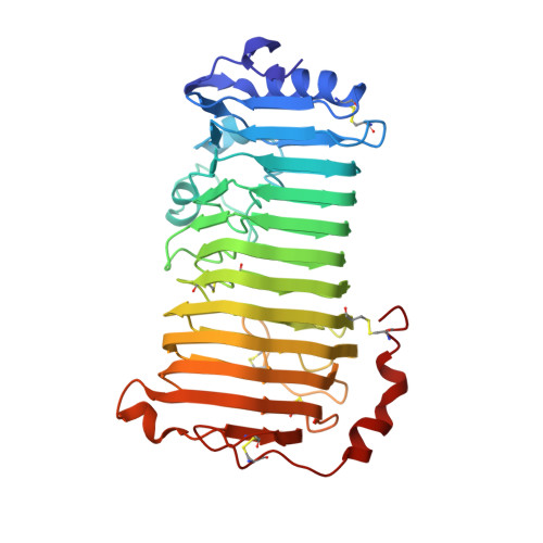
    Polygalacturonase-inhibiting proteins (PGIPs) interact with pathogen-derived polygalacturonases to inhibit their virulence-associated plant cell wall-degrading activity but stimulate immunity-inducing oligogalacturonide production. Here we show that interaction between Phaseolus vulgaris PGIP2 (PvPGIP2) and Fusarium phyllophilum polygalacturonase (FpPG) enhances substrate binding, resulting in inhibition of the enzyme activity of FpPG. This interaction promotes FpPG-catalyzed production of long-chain immunoactive oligogalacturonides, while diminishing immunosuppressive short oligogalacturonides. PvPGIP2 binding creates a substrate binding site on PvPGIP2-FpPG, forming a new polygalacturonase with boosted substrate binding activity and altered substrate preference. Structure-based engineering converts a putative PGIP that initially lacks FpPG-binding activity into an effective FpPG-interacting protein. These findings unveil a mechanism for plants to transform pathogen virulence activity into a defense trigger and provide proof of principle for engineering PGIPs with broader specificity.


  • Organizational Affiliation

    Center for Plant Biology, School of Life Sciences, Tsinghua University, Beijing 100084, China.


Macromolecules
Find similar proteins by:  (by identity cutoff)  |  3D Structure
Entity ID: 1
MoleculeChains Sequence LengthOrganismDetailsImage
Pectin lyase-like superfamily protein390Arabidopsis thalianaMutation(s): 0 
Gene Names: At5g14650AT5G14650T15N1.140T15N1_140
UniProt
Find proteins for Q9LYJ5 (Arabidopsis thaliana)
Explore Q9LYJ5 
Go to UniProtKB:  Q9LYJ5
Entity Groups  
Sequence Clusters30% Identity50% Identity70% Identity90% Identity95% Identity100% Identity
UniProt GroupQ9LYJ5
Sequence Annotations
Expand
  • Reference Sequence
Oligosaccharides

Help

Entity ID: 2
MoleculeChains Length2D Diagram Glycosylation3D Interactions
alpha-D-mannopyranose-(1-3)-[alpha-D-mannopyranose-(1-6)]alpha-D-mannopyranose-(1-6)-[alpha-D-mannopyranose-(1-3)]beta-D-mannopyranose-(1-4)-2-acetamido-2-deoxy-beta-D-glucopyranose-(1-4)-2-acetamido-2-deoxy-beta-D-glucopyranose
B
7N-Glycosylation
Glycosylation Resources
GlyTouCan:  G55220VL
GlyCosmos:  G55220VL
GlyGen:  G55220VL
Small Molecules
Ligands 1 Unique
IDChains Name / Formula / InChI Key2D Diagram3D Interactions
NAG
Query on NAG

Download Ideal Coordinates CCD File 
C [auth A]2-acetamido-2-deoxy-beta-D-glucopyranose
C8 H15 N O6
OVRNDRQMDRJTHS-FMDGEEDCSA-N
Experimental Data & Validation

Experimental Data

  • Method: X-RAY DIFFRACTION
  • Resolution: 1.50 Å
  • R-Value Free: 0.179 
  • R-Value Work: 0.158 
  • R-Value Observed: 0.159 
  • Space Group: P 1
Unit Cell:
Length ( Å )Angle ( ˚ )
a = 39.212α = 94.59
b = 43.54β = 99.99
c = 63.478γ = 110.55
Software Package:
Software NamePurpose
HKL-2000data scaling
PHENIXrefinement
HKL-2000data reduction
PHENIXphasing

Structure Validation

View Full Validation Report


Ligand Structure Quality Assessment 


Entry History & Funding Information

Deposition Data


Funding OrganizationLocationGrant Number
National Natural Science Foundation of China (NSFC)China--

Revision History  (Full details and data files)

  • Version 1.0: 2024-02-07
    Type: Initial release
  • Version 1.1: 2024-02-28
    Changes: Database references