8IHP

Structure of Semliki Forest virus VLP in complex with the receptor VLDLR-LA3


Experimental Data Snapshot

  • Method: ELECTRON MICROSCOPY
  • Resolution: 3.00 Å
  • Aggregation State: PARTICLE 
  • Reconstruction Method: SINGLE PARTICLE 

wwPDB Validation   3D Report Full Report


This is version 1.2 of the entry. See complete history


Literature

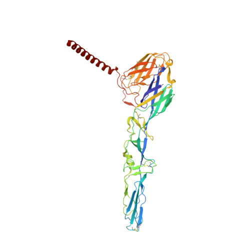
Structure of Semliki Forest virus in complex with its receptor VLDLR.

Cao, D.Ma, B.Cao, Z.Zhang, X.Xiang, Y.

(2023) Cell 186: 2208-2218.e15

  • DOI: https://doi.org/10.1016/j.cell.2023.03.032
  • Primary Citation of Related Structures:  
    8IHP

  • PubMed Abstract: 

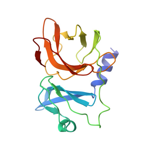
    Semliki Forest virus (SFV) is an alphavirus that uses the very-low-density lipoprotein receptor (VLDLR) as a receptor during infection of its vertebrate hosts and insect vectors. Herein, we used cryoelectron microscopy to study the structure of SFV in complex with VLDLR. We found that VLDLR binds multiple E1-DIII sites of SFV through its membrane-distal LDLR class A (LA) repeats. Among the LA repeats of the VLDLR, LA3 has the best binding affinity to SFV. The high-resolution structure shows that LA3 binds SFV E1-DIII through a small surface area of 378 Å 2 , with the main interactions at the interface involving salt bridges. Compared with the binding of single LA3s, consecutive LA repeats around LA3 promote synergistic binding to SFV, during which the LAs undergo a rotation, allowing simultaneous key interactions at multiple E1-DIII sites on the virion and enabling the binding of VLDLRs from divergent host species to SFV.


  • Organizational Affiliation

    National Laboratory of Biomacromolecules, CAS Center for Excellence in Biomacromolecules, Institute of Biophysics, Chinese Academy of Sciences (CAS), Beijing 100101, China.


Macromolecules
Find similar proteins by:  (by identity cutoff)  |  3D Structure
Entity ID: 1
MoleculeChains Sequence LengthOrganismDetailsImage
Spike glycoprotein E2
A, D, G, J
422Semliki Forest virusMutation(s): 0 
Membrane Entity: Yes 
UniProt
Find proteins for P03315 (Semliki forest virus)
Explore P03315 
Go to UniProtKB:  P03315
Entity Groups  
Sequence Clusters30% Identity50% Identity70% Identity90% Identity95% Identity100% Identity
UniProt GroupP03315
Sequence Annotations
Expand
  • Reference Sequence
Find similar proteins by:  (by identity cutoff)  |  3D Structure
Entity ID: 2
MoleculeChains Sequence LengthOrganismDetailsImage
Spike glycoprotein E1
B, E, H, K
438Semliki Forest virusMutation(s): 1 
Membrane Entity: Yes 
UniProt
Find proteins for P03315 (Semliki forest virus)
Explore P03315 
Go to UniProtKB:  P03315
Entity Groups  
Sequence Clusters30% Identity50% Identity70% Identity90% Identity95% Identity100% Identity
UniProt GroupP03315
Sequence Annotations
Expand
  • Reference Sequence
Find similar proteins by:  (by identity cutoff)  |  3D Structure
Entity ID: 3
MoleculeChains Sequence LengthOrganismDetailsImage
Capsid protein
C, F, I, L
162Semliki Forest virusMutation(s): 0 
EC: 3.4.21.90
Membrane Entity: Yes 
UniProt
Find proteins for P03315 (Semliki forest virus)
Explore P03315 
Go to UniProtKB:  P03315
Entity Groups  
Sequence Clusters30% Identity50% Identity70% Identity90% Identity95% Identity100% Identity
UniProt GroupP03315
Sequence Annotations
Expand
  • Reference Sequence
Find similar proteins by:  (by identity cutoff)  |  3D Structure
Entity ID: 4
MoleculeChains Sequence LengthOrganismDetailsImage
Very low-density lipoprotein receptor
M, N, O
39Homo sapiensMutation(s): 0 
Gene Names: VLDLR
Membrane Entity: Yes 
UniProt & NIH Common Fund Data Resources
Find proteins for P98155 (Homo sapiens)
Explore P98155 
Go to UniProtKB:  P98155
PHAROS:  P98155
GTEx:  ENSG00000147852 
Entity Groups  
Sequence Clusters30% Identity50% Identity70% Identity90% Identity95% Identity100% Identity
UniProt GroupP98155
Sequence Annotations
Expand
  • Reference Sequence
Small Molecules
Ligands 2 Unique
IDChains Name / Formula / InChI Key2D Diagram3D Interactions
NAG
Query on NAG

Download Ideal Coordinates CCD File 
AA [auth K]
P [auth A]
Q [auth A]
R [auth B]
S [auth D]
AA [auth K],
P [auth A],
Q [auth A],
R [auth B],
S [auth D],
T [auth D],
U [auth E],
V [auth G],
W [auth G],
X [auth H],
Y [auth J],
Z [auth J]
2-acetamido-2-deoxy-beta-D-glucopyranose
C8 H15 N O6
OVRNDRQMDRJTHS-FMDGEEDCSA-N
CA
Query on CA

Download Ideal Coordinates CCD File 
BA [auth M],
CA [auth N],
DA [auth O]
CALCIUM ION
Ca
BHPQYMZQTOCNFJ-UHFFFAOYSA-N
Experimental Data & Validation

Experimental Data

  • Method: ELECTRON MICROSCOPY
  • Resolution: 3.00 Å
  • Aggregation State: PARTICLE 
  • Reconstruction Method: SINGLE PARTICLE 

Structure Validation

View Full Validation Report


Entry History & Funding Information

Deposition Data


Funding OrganizationLocationGrant Number
Ministry of Science and Technology (MoST, China)China--
National Natural Science Foundation of China (NSFC)China--
Chinese Academy of SciencesChina--

Revision History  (Full details and data files)

  • Version 1.0: 2023-04-12
    Type: Initial release
  • Version 1.1: 2023-05-17
    Changes: Database references
  • Version 1.2: 2023-05-24
    Changes: Database references