8IDQ

Crystal structure of reducing-end xylose-releasing exoxylanase in GH30 from Talaromyces cellulolyticus with xylose


Experimental Data Snapshot

  • Method: X-RAY DIFFRACTION
  • Resolution: 1.70 Å
  • R-Value Free: 0.233 
  • R-Value Work: 0.199 
  • R-Value Observed: 0.201 

wwPDB Validation   3D Report Full Report


Ligand Structure Quality Assessment 


This is version 1.1 of the entry. See complete history


Literature

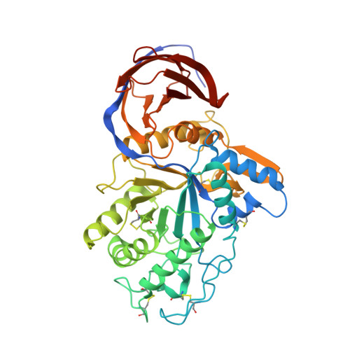
Crystal structure of reducing-end xylose-releasing exoxylanase in subfamily 7 of glycoside hydrolase family 30.

Nakamichi, Y.Watanabe, M.Fujii, T.Inoue, H.Morita, T.

(2023) Proteins 91: 1341-1350

  • DOI: https://doi.org/10.1002/prot.26505
  • Primary Citation of Related Structures:  
    8IDP, 8IDQ

  • PubMed Abstract: 

    TcXyn30A from Talaromyces cellulolyticus, which belongs to subfamily 7 of the glycoside hydrolase family 30 (GH30-7), releases xylose from the reducing end of xylan and xylooligosaccharides (XOSs), the so-called reducing-end xylose-releasing exoxylanase (ReX). In this study, the crystal structures of TcXyn30A with and without xylose at subsite +1 (the binding site of the xylose residue at the reducing end) were determined. This is the first report on the structure of ReX in the family GH30-7. TcXyn30A forms a dimer. The complex structure of TcXyn30A with xylose revealed that subsite +1 is located at the dimer interface. TcXyn30A recognizes xylose at subsite +1 composed of amino acid residues from each monomer and blocks substrate binding to subsite +2 by dimer formation. Thus, the dimeric conformation is responsible for ReX activity. The structural comparison between TcXyn30A and the homologous enzyme indicated that subsite -2 is composed of assembled three stacked Trp residues, Trp49, Trp333, and Trp334, allowing TcXyn30A to accommodate xylan and any branched XOSs decorated with a substitution such as α-1,2-linked 4-O-methyl-d-glucuronic acid or α-1,2- and/or -1,3-linked L-arabinofuranose. These findings provide an insight into the structural determinants for ReX activity of TcXyn30A.


  • Organizational Affiliation

    Research Institute for Sustainable Chemistry, National Institute of Advanced Industrial Science and Technology (AIST), Higashi-Hiroshima, Hiroshima, Japan.


Macromolecules
Find similar proteins by:  (by identity cutoff)  |  3D Structure
Entity ID: 1
MoleculeChains Sequence LengthOrganismDetailsImage
Reducing-end xylose-releasing exoxylanase Xyn30A
A, B, C, D
462Talaromyces pinophilusMutation(s): 0 
Gene Names: TCE0_047r17950
UniProt
Find proteins for A0A0B8MZ29 (Talaromyces pinophilus)
Explore A0A0B8MZ29 
Go to UniProtKB:  A0A0B8MZ29
Entity Groups  
Sequence Clusters30% Identity50% Identity70% Identity90% Identity95% Identity100% Identity
UniProt GroupA0A0B8MZ29
Sequence Annotations
Expand
  • Reference Sequence
Oligosaccharides

Help

Entity ID: 2
MoleculeChains Length2D Diagram Glycosylation3D Interactions
alpha-D-mannopyranose-(1-2)-alpha-D-mannopyranose-(1-2)-alpha-D-mannopyranose-(1-3)-[alpha-D-mannopyranose-(1-2)-alpha-D-mannopyranose-(1-3)-[alpha-D-mannopyranose-(1-2)-alpha-D-mannopyranose-(1-6)]alpha-D-mannopyranose-(1-6)]beta-D-mannopyranose-(1-4)-2-acetamido-2-deoxy-beta-D-glucopyranose-(1-4)-2-acetamido-2-deoxy-beta-D-glucopyranose
E, N
11N-Glycosylation
Glycosylation Resources
GlyTouCan:  G60230HH
GlyCosmos:  G60230HH
GlyGen:  G60230HH
Entity ID: 3
MoleculeChains Length2D Diagram Glycosylation3D Interactions
2-acetamido-2-deoxy-beta-D-glucopyranose-(1-4)-2-acetamido-2-deoxy-beta-D-glucopyranose
F, J, M, O
2N-Glycosylation
Glycosylation Resources
GlyTouCan:  G42666HT
GlyCosmos:  G42666HT
GlyGen:  G42666HT
Entity ID: 4
MoleculeChains Length2D Diagram Glycosylation3D Interactions
alpha-D-mannopyranose-(1-3)-alpha-D-mannopyranose-(1-6)-[alpha-D-mannopyranose-(1-3)]beta-D-mannopyranose-(1-4)-2-acetamido-2-deoxy-beta-D-glucopyranose-(1-4)-2-acetamido-2-deoxy-beta-D-glucopyranose
G, I, P
6N-Glycosylation
Glycosylation Resources
GlyTouCan:  G09724ZC
GlyCosmos:  G09724ZC
GlyGen:  G09724ZC
Entity ID: 5
MoleculeChains Length2D Diagram Glycosylation3D Interactions
alpha-D-mannopyranose-(1-2)-alpha-D-mannopyranose-(1-3)-[alpha-D-mannopyranose-(1-2)-alpha-D-mannopyranose-(1-6)]alpha-D-mannopyranose-(1-6)-[alpha-D-mannopyranose-(1-2)-alpha-D-mannopyranose-(1-3)]beta-D-mannopyranose-(1-4)-2-acetamido-2-deoxy-beta-D-glucopyranose-(1-4)-2-acetamido-2-deoxy-beta-D-glucopyranose
H
10N-Glycosylation
Glycosylation Resources
GlyTouCan:  G89864BN
GlyCosmos:  G89864BN
GlyGen:  G89864BN
Entity ID: 6
MoleculeChains Length2D Diagram Glycosylation3D Interactions
alpha-D-mannopyranose-(1-2)-alpha-D-mannopyranose-(1-6)-[alpha-D-mannopyranose-(1-3)]alpha-D-mannopyranose-(1-6)-[alpha-D-mannopyranose-(1-2)-alpha-D-mannopyranose-(1-3)]beta-D-mannopyranose-(1-4)-2-acetamido-2-deoxy-beta-D-glucopyranose-(1-4)-2-acetamido-2-deoxy-beta-D-glucopyranose
K
9N-Glycosylation
Glycosylation Resources
GlyTouCan:  G83161QT
GlyCosmos:  G83161QT
GlyGen:  G83161QT
Entity ID: 7
MoleculeChains Length2D Diagram Glycosylation3D Interactions
alpha-D-mannopyranose-(1-3)-[alpha-D-mannopyranose-(1-6)]beta-D-mannopyranose-(1-4)-2-acetamido-2-deoxy-beta-D-glucopyranose-(1-4)-2-acetamido-2-deoxy-beta-D-glucopyranose
L
5N-Glycosylation
Glycosylation Resources
GlyTouCan:  G22768VO
GlyCosmos:  G22768VO
GlyGen:  G22768VO
Small Molecules
Ligands 4 Unique
IDChains Name / Formula / InChI Key2D Diagram3D Interactions
NAG
Query on NAG

Download Ideal Coordinates CCD File 
BA [auth C]
CA [auth C]
GA [auth D]
IA [auth D]
Q [auth A]
BA [auth C],
CA [auth C],
GA [auth D],
IA [auth D],
Q [auth A],
R [auth A],
U [auth B],
W [auth B]
2-acetamido-2-deoxy-beta-D-glucopyranose
C8 H15 N O6
OVRNDRQMDRJTHS-FMDGEEDCSA-N
XYP (Subject of Investigation/LOI)
Query on XYP

Download Ideal Coordinates CCD File 
EA [auth C],
LA [auth D],
T [auth A],
Z [auth B]
beta-D-xylopyranose
C5 H10 O5
SRBFZHDQGSBBOR-KKQCNMDGSA-N
GOL
Query on GOL

Download Ideal Coordinates CCD File 
DA [auth C]
HA [auth D]
JA [auth D]
KA [auth D]
S [auth A]
DA [auth C],
HA [auth D],
JA [auth D],
KA [auth D],
S [auth A],
V [auth B],
X [auth B],
Y [auth B]
GLYCEROL
C3 H8 O3
PEDCQBHIVMGVHV-UHFFFAOYSA-N
CL
Query on CL

Download Ideal Coordinates CCD File 
AA [auth B],
FA [auth C]
CHLORIDE ION
Cl
VEXZGXHMUGYJMC-UHFFFAOYSA-M
Experimental Data & Validation

Experimental Data

  • Method: X-RAY DIFFRACTION
  • Resolution: 1.70 Å
  • R-Value Free: 0.233 
  • R-Value Work: 0.199 
  • R-Value Observed: 0.201 
  • Space Group: P 21 21 2
Unit Cell:
Length ( Å )Angle ( ˚ )
a = 138.249α = 90
b = 120.159β = 90
c = 122.886γ = 90
Software Package:
Software NamePurpose
XDSdata scaling
XDSdata reduction
MOLREPphasing
PHENIXrefinement

Structure Validation

View Full Validation Report


Ligand Structure Quality Assessment 


Entry History & Funding Information

Deposition Data


Funding OrganizationLocationGrant Number
Not funded--

Revision History  (Full details and data files)

  • Version 1.0: 2023-05-17
    Type: Initial release
  • Version 1.1: 2023-08-23
    Changes: Data collection, Database references