8I1V

The asymmetric unit of P22 procapsid


Experimental Data Snapshot

  • Method: ELECTRON MICROSCOPY
  • Resolution: 2.60 Å
  • Aggregation State: PARTICLE 
  • Reconstruction Method: SINGLE PARTICLE 

wwPDB Validation   3D Report Full Report


This is version 1.1 of the entry. See complete history


Literature

Assembly and Capsid Expansion Mechanism of Bacteriophage P22 Revealed by High-Resolution Cryo-EM Structures.

Xiao, H.Zhou, J.Yang, F.Liu, Z.Song, J.Chen, W.Liu, H.Cheng, L.

(2023) Viruses 15

  • DOI: https://doi.org/10.3390/v15020355
  • Primary Citation of Related Structures:  
    8I1T, 8I1V

  • PubMed Abstract: 

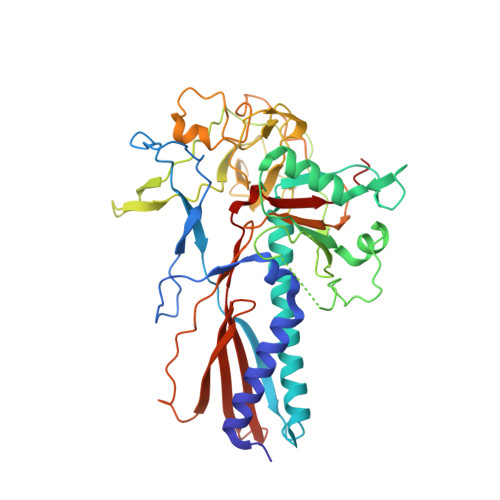
    The formation of many double-stranded DNA viruses, such as herpesviruses and bacteriophages, begins with the scaffolding-protein-mediated assembly of the procapsid. Subsequently, the procapsid undergoes extensive structural rearrangement and expansion to become the mature capsid. Bacteriophage P22 is an established model system used to study virus maturation. Here, we report the cryo-electron microscopy structures of procapsid, empty procapsid, empty mature capsid, and mature capsid of phage P22 at resolutions of 2.6 Å, 3.9 Å, 2.8 Å, and 3.0 Å, respectively. The structure of the procapsid allowed us to build an accurate model of the coat protein gp5 and the C-terminal region of the scaffolding protein gp8. In addition, interactions among the gp5 subunits responsible for procapsid assembly and stabilization were identified. Two C-terminal α-helices of gp8 were observed to interact with the coat protein in the procapsid. The amino acid interactions between gp5 and gp8 in the procapsid were consistent with the results of previous biochemical studies involving mutant proteins. Our structures reveal hydrogen bonds and salt bridges between the gp5 subunits in the procapsid and the conformational changes of the gp5 domains involved in the closure of the local sixfold opening and a thinner capsid shell during capsid maturation.


  • Organizational Affiliation

    Institute of Interdisciplinary Studies, Key Laboratory for Matter Microstructure and Function of Hunan Province, Key Laboratory of Low-dimensional Quantum Structures and Quantum Control, Hunan Normal University, Changsha 410082, China.


Macromolecules
Find similar proteins by:  (by identity cutoff)  |  3D Structure
Entity ID: 1
MoleculeChains Sequence LengthOrganismDetailsImage
Major capsid protein430Salmonella phage P22Mutation(s): 0 
UniProt
Find proteins for P26747 (Salmonella phage P22)
Explore P26747 
Go to UniProtKB:  P26747
Entity Groups  
Sequence Clusters30% Identity50% Identity70% Identity90% Identity95% Identity100% Identity
UniProt GroupP26747
Sequence Annotations
Expand
  • Reference Sequence
Find similar proteins by:  (by identity cutoff)  |  3D Structure
Entity ID: 2
MoleculeChains Sequence LengthOrganismDetailsImage
Scaffolding protein
H, I, J, K, L
H, I, J, K, L, M, N
303Salmonella phage P22Mutation(s): 0 
UniProt
Find proteins for P26748 (Salmonella phage P22)
Explore P26748 
Go to UniProtKB:  P26748
Entity Groups  
Sequence Clusters30% Identity50% Identity70% Identity90% Identity95% Identity100% Identity
UniProt GroupP26748
Sequence Annotations
Expand
  • Reference Sequence
Experimental Data & Validation

Experimental Data

  • Method: ELECTRON MICROSCOPY
  • Resolution: 2.60 Å
  • Aggregation State: PARTICLE 
  • Reconstruction Method: SINGLE PARTICLE 

Structure Validation

View Full Validation Report


Entry History & Funding Information

Deposition Data


Funding OrganizationLocationGrant Number
National Natural Science Foundation of China (NSFC)China12034006
National Natural Science Foundation of China (NSFC)China31971122
National Natural Science Foundation of China (NSFC)China32071209
National Natural Science Foundation of China (NSFC)China32200994

Revision History  (Full details and data files)

  • Version 1.0: 2023-03-08
    Type: Initial release
  • Version 1.1: 2023-03-15
    Changes: Database references