8HTY

Candida boidinii Formate Dehydrogenase Crystal Structure at 1.4 Angstrom Resolution


Experimental Data Snapshot

  • Method: X-RAY DIFFRACTION
  • Resolution: 1.40 Å
  • R-Value Free: 0.190 
  • R-Value Work: 0.167 
  • R-Value Observed: 0.168 

wwPDB Validation   3D Report Full Report


This is version 1.1 of the entry. See complete history


Literature

Structural analysis of wild-type and Val120Thr mutant Candida boidinii formate dehydrogenase by X-ray crystallography.

Gul, M.Yuksel, B.Bulut, H.DeMirci, H.

(2023) Acta Crystallogr D Struct Biol 79: 1010-1017

  • DOI: https://doi.org/10.1107/S2059798323008070
  • Primary Citation of Related Structures:  
    8HTY, 8IQ7, 8IVJ

  • PubMed Abstract: 

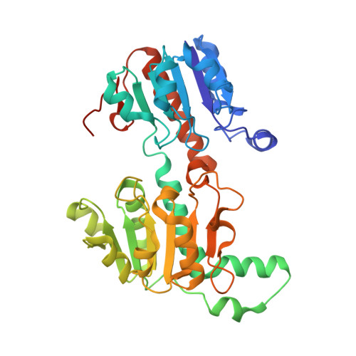
    Candida boidinii NAD + -dependent formate dehydrogenase (CbFDH) has gained significant attention for its potential application in the production of biofuels and various industrial chemicals from inorganic carbon dioxide. The present study reports the atomic X-ray crystal structures of wild-type CbFDH at cryogenic and ambient temperatures, as well as that of the Val120Thr mutant at cryogenic temperature, determined at the Turkish Light Source `Turkish DeLight'. The structures reveal new hydrogen bonds between Thr120 and water molecules in the active site of the mutant CbFDH, suggesting increased stability of the active site and more efficient electron transfer during the reaction. Further experimental data is needed to test these hypotheses. Collectively, these findings provide invaluable insights into future protein-engineering efforts that could potentially enhance the efficiency and effectiveness of CbFDH.


  • Organizational Affiliation

    Department of Molecular Biology and Genetics, Koc University, 34450 Istanbul, Türkiye.


Macromolecules
Find similar proteins by:  (by identity cutoff)  |  3D Structure
Entity ID: 1
MoleculeChains Sequence LengthOrganismDetailsImage
Formate dehydrogenase
A, B, C, D
364[Candida] boidiniiMutation(s): 0 
Gene Names: FDH
UniProt
Find proteins for A0A0A1EQY0 (Candida boidinii)
Explore A0A0A1EQY0 
Go to UniProtKB:  A0A0A1EQY0
Entity Groups  
Sequence Clusters30% Identity50% Identity70% Identity90% Identity95% Identity100% Identity
UniProt GroupA0A0A1EQY0
Sequence Annotations
Expand
  • Reference Sequence
Small Molecules
Ligands 1 Unique
IDChains Name / Formula / InChI Key2D Diagram3D Interactions
SO4
Query on SO4

Download Ideal Coordinates CCD File 
E [auth A]
F [auth A]
G [auth A]
H [auth A]
I [auth A]
E [auth A],
F [auth A],
G [auth A],
H [auth A],
I [auth A],
J [auth A],
K [auth B],
L [auth B],
M [auth B],
N [auth B],
O [auth C],
P [auth C],
Q [auth D],
R [auth D]
SULFATE ION
O4 S
QAOWNCQODCNURD-UHFFFAOYSA-L
Experimental Data & Validation

Experimental Data

  • Method: X-RAY DIFFRACTION
  • Resolution: 1.40 Å
  • R-Value Free: 0.190 
  • R-Value Work: 0.167 
  • R-Value Observed: 0.168 
  • Space Group: P 1
Unit Cell:
Length ( Å )Angle ( ˚ )
a = 54.132α = 78.14
b = 68.959β = 89.48
c = 109.792γ = 81.11
Software Package:
Software NamePurpose
PHENIXrefinement
PHASERphasing
CrysalisProdata reduction
CrysalisProdata scaling

Structure Validation

View Full Validation Report


Entry History & Funding Information

Deposition Data


Funding OrganizationLocationGrant Number
Not funded--

Revision History  (Full details and data files)

  • Version 1.0: 2023-01-18
    Type: Initial release
  • Version 1.1: 2023-11-08
    Changes: Data collection, Database references