


Improved thermostability of a glucose-tolerant glycosidase based on its X-ray crystal structure
Dong, P.P., Wu, Y.K.To be published.
Experimental Data Snapshot
wwPDB Validation 3D Report Full Report

Entity ID: 1 | |||||
|---|---|---|---|---|---|
| Molecule | Chains | Sequence Length | Organism | Details | Image |
| Beta-glucosidase | 447 | Caldibacillus thermoamylovorans | Mutation(s): 0 Gene Names: CQJ30_16875 EC: 3.2.1.21 | 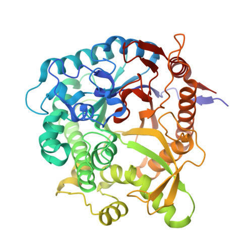 | |
UniProt | |||||
Find proteins for A0A2U8EHQ1 (Caldibacillus thermoamylovorans) Explore A0A2U8EHQ1 Go to UniProtKB: A0A2U8EHQ1 | |||||
Entity Groups | |||||
| Sequence Clusters | 30% Identity50% Identity70% Identity90% Identity95% Identity100% Identity | ||||
| UniProt Group | A0A2U8EHQ1 | ||||
Sequence AnnotationsExpand | |||||
| |||||
| Length ( Å ) | Angle ( ˚ ) |
|---|---|
| a = 166.3 | α = 90 |
| b = 166.3 | β = 90 |
| c = 122.41 | γ = 90 |
| Software Name | Purpose |
|---|---|
| PHENIX | refinement |
| HKL-2000 | data reduction |
| Aimless | data scaling |
| PHENIX | phasing |
| Funding Organization | Location | Grant Number |
|---|---|---|
| National Natural Science Foundation of China (NSFC) | China | -- |
RCSB PDB (citation) is hosted by
RCSB PDB is a member of the

