


crystal structure of UGT71AP2 in complex with UDP
Wang, Z.L., He, C., Li, F., Qiao, X., Ye, M.To be published.
Experimental Data Snapshot
Entity ID: 1 | |||||
|---|---|---|---|---|---|
| Molecule | Chains | Sequence Length | Organism | Details | Image |
| UGT71AP2 | 464 | Scutellaria baicalensis | Mutation(s): 0 | 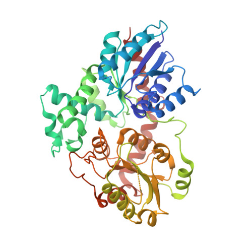 | |
Entity Groups | |||||
| Sequence Clusters | 30% Identity50% Identity70% Identity90% Identity95% Identity100% Identity | ||||
Sequence AnnotationsExpand | |||||
| |||||
| Ligands 1 Unique | |||||
|---|---|---|---|---|---|
| ID | Chains | Name / Formula / InChI Key | 2D Diagram | 3D Interactions | |
| UDP (Subject of Investigation/LOI) Query on UDP | C [auth A], D [auth B] | URIDINE-5'-DIPHOSPHATE C9 H14 N2 O12 P2 XCCTYIAWTASOJW-XVFCMESISA-N |  | ||
| Length ( Å ) | Angle ( ˚ ) |
|---|---|
| a = 104.728 | α = 90 |
| b = 108.544 | β = 90 |
| c = 84.35 | γ = 90 |
| Software Name | Purpose |
|---|---|
| HKL-2000 | data scaling |
| PHENIX | refinement |
| PDB_EXTRACT | data extraction |
| DENZO | data reduction |
| PHASES | phasing |
| Funding Organization | Location | Grant Number |
|---|---|---|
| National Natural Science Foundation of China (NSFC) | China | -- |
RCSB PDB (citation) is hosted by
RCSB PDB is a member of the

