Aspergillus oryzae alpha-l-rhamnosidase: Crystal structure and insight into the substrate specificity.
Makabe, K., Ishida, N., Kanezaki, N., Shiono, Y., Koseki, T.(2024) Proteins 92: 236-245
- PubMed: 37818702
- DOI: https://doi.org/10.1002/prot.26608
- Primary Citation of Related Structures:
8HMM - PubMed Abstract:
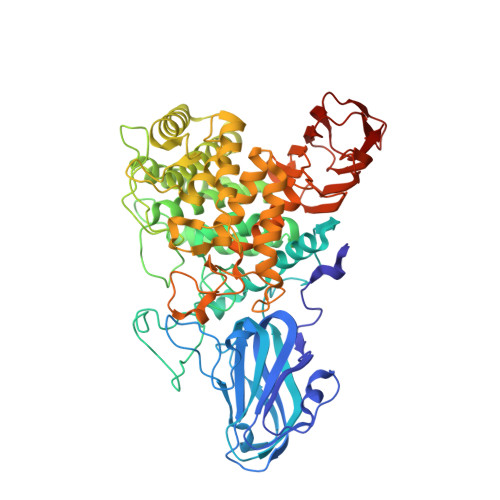
The subsequent biochemical and structural investigations of the purified recombinant α-l-rhamnosidase from Aspergillus oryzae expressed in Pichia pastoris, designated as rAoRhaA, were performed. The specific activity of the rAoRhaA wild-type was higher toward hesperidin and narirutin, where the l-rhamnose residue was α-1,6-linked to β-d-glucoside, than toward neohesperidin and naringin with an α-1,2-linkage to β-d-glucoside. However, no activity was detected toward quercitrin, myricitrin, and epimedin C. rAoRhaA kinetic analysis indicated that K m values for neohesperidin, naringin, and rutin were lower compared to those for hesperidin and narirutin. k cat values for hesperidin and narirutin were higher than those for neohesperidin, naringin, and rutin. High catalytic efficiency (k cat /K m ) toward hesperidin and narirutin was a result of a considerably high k cat value, while K m values for hesperidin and narirutin were higher than those for naringin, neohesperidin, and rutin. The crystal structure of rAoRhaA revealed that the catalytic domain was represented by an (α/α) 6 -barrel with the active site located in a deep cleft and two β-sheet domains were also present in the N- and C-terminal sites of the catalytic domain. Additionally, five asparagine-attached N-acetylglucosamine molecules were observed. The catalytic residues of AoRhaA were suggested to be Asp254 and Glu524, and their catalytic roles were confirmed by mutational studies of D254N and E524Q variants, which lost their activity completely. Notably, three aspartic acids (Asp117, Asp249, and Asp261) located at the catalytic pocket were replaced with asparagine. D117N variant showed reduced activity. D249N and D261N variants activities drastically decreased.
Organizational Affiliation:
Graduate School of Science and Engineering, Faculty of Engineering, Yamagata University, Yonezawa, Japan.