


Crystal structure of rabies virus (strain CVS-11) P3 dimerization domain
Donnelly, C.M., Cross, E.M., Forwood, J.K.To be published.
Experimental Data Snapshot
wwPDB Validation 3D Report Full Report

Entity ID: 1 | |||||
|---|---|---|---|---|---|
| Molecule | Chains | Sequence Length | Organism | Details | Image |
| Phosphoprotein | 119 | Lyssavirus rabies | Mutation(s): 0 | 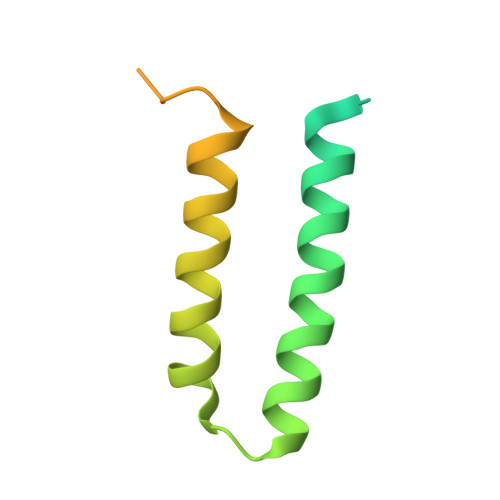 | |
UniProt | |||||
Find proteins for P22363 (Rabies virus (strain CVS-11)) Explore P22363 Go to UniProtKB: P22363 | |||||
Entity Groups | |||||
| Sequence Clusters | 30% Identity50% Identity70% Identity90% Identity95% Identity100% Identity | ||||
| UniProt Group | P22363 | ||||
Sequence AnnotationsExpand | |||||
| |||||
| Length ( Å ) | Angle ( ˚ ) |
|---|---|
| a = 67.05 | α = 90 |
| b = 44.097 | β = 100.44 |
| c = 111.787 | γ = 90 |
| Software Name | Purpose |
|---|---|
| PHASER | phasing |
| PHENIX | refinement |
| DIALS | data reduction |
| Aimless | data scaling |
| Coot | model building |
| Funding Organization | Location | Grant Number |
|---|---|---|
| Not funded | -- |
RCSB PDB (citation) is hosted by
RCSB PDB is a member of the

