8FIG

Room-temperature X-ray structure of SARS-CoV-2 main protease double mutant E290A/R298A in complex with GC373


Experimental Data Snapshot

  • Method: X-RAY DIFFRACTION
  • Resolution: 1.75 Å
  • R-Value Free: 0.185 
  • R-Value Work: 0.165 
  • R-Value Observed: 0.166 

wwPDB Validation   3D Report Full Report


Ligand Structure Quality Assessment 


This is version 1.0 of the entry. See complete history


Literature

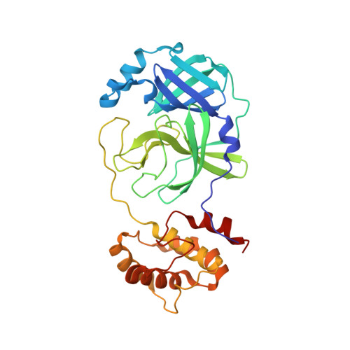
Insights into the mechanism of SARS-CoV-2 main protease autocatalytic maturation from model precursors.

Aniana, A.Nashed, N.T.Ghirlando, R.Coates, L.Kneller, D.W.Kovalevsky, A.Louis, J.M.

(2023) Commun Biol 6: 1159-1159

  • DOI: https://doi.org/10.1038/s42003-023-05469-8
  • Primary Citation of Related Structures:  
    8FIG

  • PubMed Abstract: 

    A critical step for SARS-CoV-2 assembly and maturation involves the autoactivation of the main protease (MPro WT ) from precursor polyproteins. Upon expression, a model precursor of MPro WT mediates its own release at its termini rapidly to yield a mature dimer. A construct with an E290A mutation within MPro exhibits time dependent autoprocessing of the accumulated precursor at the N-terminal nsp4/nsp5 site followed by the C-terminal nsp5/nsp6 cleavage. In contrast, a precursor containing E290A and R298A mutations (MPro M ) displays cleavage only at the nsp4/nsp5 site to yield an intermediate monomeric product, which is cleaved at the nsp5/nsp6 site only by MPro WT . MPro M and the catalytic domain (MPro 1-199 ) fused to the truncated nsp4 region also show time-dependent conversion in vitro to produce MPro M and MPro 1-199 , respectively. The reactions follow first-order kinetics indicating that the nsp4/nsp5 cleavage occurs via an intramolecular mechanism. These results support a mechanism involving an N-terminal intramolecular cleavage leading to an increase in the dimer population and followed by an intermolecular cleavage at the C-terminus. Thus, targeting the predominantly monomeric MPro precursor for inhibition may lead to the identification of potent drugs for treatment.


  • Organizational Affiliation

    Laboratory of Chemical Physics, National Institute of Diabetes and Digestive and Kidney Diseases, National Institutes of Health, DHHS, Bethesda, MD, 20892-0520, USA.


Macromolecules
Find similar proteins by:  (by identity cutoff)  |  3D Structure
Entity ID: 1
MoleculeChains Sequence LengthOrganismDetailsImage
3C-like proteinase nsp5306Severe acute respiratory syndrome coronavirus 2Mutation(s): 2 
UniProt
Find proteins for P0DTD1 (Severe acute respiratory syndrome coronavirus 2)
Explore P0DTD1 
Go to UniProtKB:  P0DTD1
Entity Groups  
Sequence Clusters30% Identity50% Identity70% Identity90% Identity95% Identity100% Identity
UniProt GroupP0DTD1
Sequence Annotations
Expand
  • Reference Sequence
Small Molecules
Ligands 1 Unique
IDChains Name / Formula / InChI Key2D Diagram3D Interactions
UED (Subject of Investigation/LOI)
Query on UED

Download Ideal Coordinates CCD File 
B [auth A]N~2~-[(benzyloxy)carbonyl]-N-{(2S)-1-hydroxy-3-[(3S)-2-oxopyrrolidin-3-yl]propan-2-yl}-L-leucinamide
C21 H31 N3 O5
JUCVXDDMQHPCKT-BZSNNMDCSA-N
Experimental Data & Validation

Experimental Data

  • Method: X-RAY DIFFRACTION
  • Resolution: 1.75 Å
  • R-Value Free: 0.185 
  • R-Value Work: 0.165 
  • R-Value Observed: 0.166 
  • Space Group: I 1 2 1
Unit Cell:
Length ( Å )Angle ( ˚ )
a = 52.243α = 90
b = 80.73β = 95.65
c = 92.547γ = 90
Software Package:
Software NamePurpose
PHENIXrefinement
CrysalisProdata reduction
CrysalisProdata scaling
PHASERphasing

Structure Validation

View Full Validation Report


Ligand Structure Quality Assessment 


Entry History & Funding Information

Deposition Data


Funding OrganizationLocationGrant Number
Not funded--

Revision History  (Full details and data files)

  • Version 1.0: 2023-11-22
    Type: Initial release