8FFC

Crystal structure of iron bound Dps protein (PA0962) from Pseudomonas aeruginosa (cubic form)


Experimental Data Snapshot

  • Method: X-RAY DIFFRACTION
  • Resolution: 1.85 Å
  • R-Value Free: 0.192 
  • R-Value Work: 0.162 
  • R-Value Observed: 0.163 

wwPDB Validation   3D Report Full Report


This is version 1.1 of the entry. See complete history


Literature

Pseudomonas aeruginosa Dps (PA0962) Functions in H 2 O 2 Mediated Oxidative Stress Defense and Exhibits In Vitro DNA Cleaving Activity.

Rajapaksha, N.Soldano, A.Yao, H.Donnarumma, F.Kashipathy, M.M.Seibold, S.Battaile, K.P.Lovell, S.Rivera, M.

(2023) Int J Mol Sci 24

  • DOI: https://doi.org/10.3390/ijms24054669
  • Primary Citation of Related Structures:  
    8FF9, 8FFA, 8FFB, 8FFC, 8FFD

  • PubMed Abstract: 

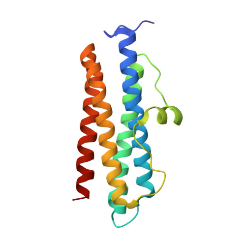
    We report the structural, biochemical, and functional characterization of the product of gene PA0962 from Pseudomonas aeruginosa PAO1. The protein, termed Pa Dps, adopts the Dps subunit fold and oligomerizes into a nearly spherical 12-mer quaternary structure at pH 6.0 or in the presence of divalent cations at neutral pH and above. The 12-Mer Pa Dps contains two di-iron centers at the interface of each subunit dimer, coordinated by conserved His, Glu, and Asp residues. In vitro, the di-iron centers catalyze the oxidation of Fe 2+ utilizing H 2 O 2 (not O 2 ) as an oxidant, suggesting Pa Dps functions to aid P. aeruginosa to survive H 2 O 2 -mediated oxidative stress. In agreement, a P. aeruginosa Δ dps mutant is significantly more susceptible to H 2 O 2 than the parent strain. The Pa Dps structure harbors a novel network of Tyr residues at the interface of each subunit dimer between the two di-iron centers, which captures radicals generated during Fe 2+ oxidation at the ferroxidase centers and forms di-tyrosine linkages, thus effectively trapping the radicals within the Dps shell. Surprisingly, incubating Pa Dps and DNA revealed unprecedented DNA cleaving activity that is independent of H 2 O 2 or O 2 but requires divalent cations and 12-mer Pa Dps.


  • Organizational Affiliation

    Department of Chemistry, Louisiana State University, 232 Choppin Hall, Baton Rouge, LA 70803, USA.


Macromolecules
Find similar proteins by:  (by identity cutoff)  |  3D Structure
Entity ID: 1
MoleculeChains Sequence LengthOrganismDetailsImage
Probable dna-binding stress protein
A, B, C, D, E
A, B, C, D, E, F, G, H, I, J, K, L, M, N, O, P
156Pseudomonas aeruginosa PAO1Mutation(s): 0 
Gene Names: PA0962
UniProt
Find proteins for Q9I4Z7 (Pseudomonas aeruginosa (strain ATCC 15692 / DSM 22644 / CIP 104116 / JCM 14847 / LMG 12228 / 1C / PRS 101 / PAO1))
Explore Q9I4Z7 
Go to UniProtKB:  Q9I4Z7
Entity Groups  
Sequence Clusters30% Identity50% Identity70% Identity90% Identity95% Identity100% Identity
UniProt GroupQ9I4Z7
Sequence Annotations
Expand
  • Reference Sequence
Small Molecules
Ligands 2 Unique
IDChains Name / Formula / InChI Key2D Diagram3D Interactions
EPE
Query on EPE

Download Ideal Coordinates CCD File 
EC [auth O]
HA [auth C]
RA [auth E]
U [auth A]
VA [auth F]
EC [auth O],
HA [auth C],
RA [auth E],
U [auth A],
VA [auth F],
VB [auth M]
4-(2-HYDROXYETHYL)-1-PIPERAZINE ETHANESULFONIC ACID
C8 H18 N2 O4 S
JKMHFZQWWAIEOD-UHFFFAOYSA-N
FE2 (Subject of Investigation/LOI)
Query on FE2

Download Ideal Coordinates CCD File 
AA [auth B]
AB [auth H]
AC [auth O]
BA [auth B]
BB [auth H]
AA [auth B],
AB [auth H],
AC [auth O],
BA [auth B],
BB [auth H],
BC [auth O],
CA [auth B],
CB [auth H],
CC [auth O],
DA [auth C],
DB [auth H],
DC [auth O],
EA [auth C],
EB [auth I],
FA [auth C],
FB [auth I],
FC [auth P],
GA [auth C],
GB [auth I],
GC [auth P],
HB [auth I],
IA [auth D],
IB [auth J],
JA [auth D],
JB [auth J],
KA [auth D],
KB [auth K],
LA [auth D],
LB [auth K],
MA [auth E],
MB [auth K],
NA [auth E],
NB [auth L],
OA [auth E],
OB [auth L],
PA [auth E],
PB [auth M],
Q [auth A],
QA [auth E],
QB [auth M],
R [auth A],
RB [auth M],
S [auth A],
SA [auth F],
SB [auth M],
T [auth A],
TA [auth F],
TB [auth M],
UA [auth F],
UB [auth M],
V [auth B],
W [auth B],
WA [auth G],
WB [auth N],
X [auth B],
XA [auth G],
XB [auth N],
Y [auth B],
YA [auth G],
YB [auth O],
Z [auth B],
ZA [auth G],
ZB [auth O]
FE (II) ION
Fe
CWYNVVGOOAEACU-UHFFFAOYSA-N
Experimental Data & Validation

Experimental Data

  • Method: X-RAY DIFFRACTION
  • Resolution: 1.85 Å
  • R-Value Free: 0.192 
  • R-Value Work: 0.162 
  • R-Value Observed: 0.163 
  • Space Group: P 21 3
Unit Cell:
Length ( Å )Angle ( ˚ )
a = 223.9α = 90
b = 223.9β = 90
c = 223.9γ = 90
Software Package:
Software NamePurpose
PHENIXrefinement
Aimlessdata scaling
XDSdata reduction
PHASERphasing

Structure Validation

View Full Validation Report


Entry History & Funding Information

Deposition Data


Funding OrganizationLocationGrant Number
National Institutes of Health/National Institute Of Allergy and Infectious Diseases (NIH/NIAID)United StatesR01AI169344
National Institutes of Health/Office of the DirectorUnited StatesS10OD030394

Revision History  (Full details and data files)

  • Version 1.0: 2023-03-08
    Type: Initial release
  • Version 1.1: 2023-03-22
    Changes: Database references