


Electrochemical and Structural Study of the Buried Tryptophan in Azurin: Effects of Hydration and Polarity on the Redox Potential of W48.
Tyson, K., Tangtartharakul, C.B., Zeug, M., Findling, N., Haddy, A., Hvastkovs, E., Choe, J.Y., Kim, J.E., Offenbacher, A.R.(2023) J Phys Chem B 127: 133-143
- PubMed: 36542812
- DOI: https://doi.org/10.1021/acs.jpcb.2c06677
- Primary Citation of Related Structures:
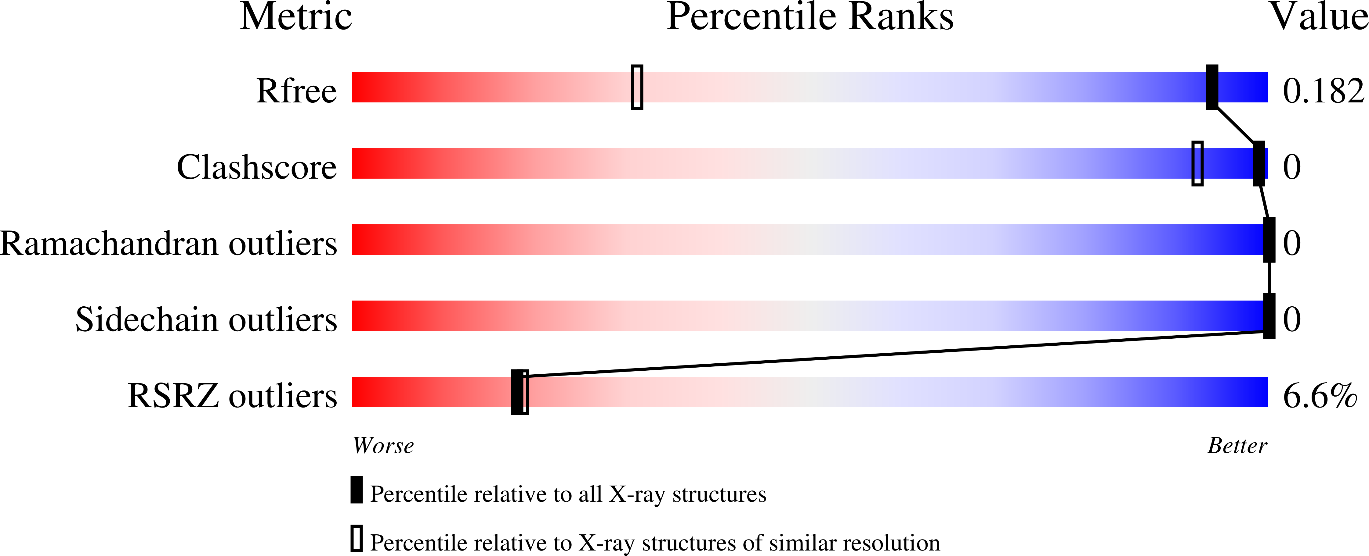
8F5K, 8F5L - PubMed Abstract:
Tryptophan serves as an important redox-active amino acid in mediating electron transfer and mitigating oxidative damage in proteins. We previously showed a difference in electrochemical potentials for two tryptophan residues in azurin with distinct hydrogen-bonding environments. Here, we test whether reducing the side chain bulk at position Phe110 to Leu, Ser, or Ala impacts the electrochemical potentials ( E °) for tryptophan at position 48. X-ray diffraction confirmed the influx of crystallographically resolved water molecules for both the F110A and F110L tyrosine free azurin mutants. The local environments of W48 in all azurin mutants were further evaluated by UV resonance Raman (UVRR) spectroscopy to probe the impact of mutations on hydrogen bonding and polarity. A correlation between the frequency of the ω17 mode─considered a vibrational marker for hydrogen bonding─and E ° is proposed. However, the trend is opposite to the expectation from a previous study on small molecules. Density functional theory calculations suggest that the ω17 mode reflects hydrogen bonding as well as local polarity. Further, the UVRR data reveal different intensity/frequency shifts of the ω9/ω10 vibrational modes that characterize the local H-bonding environments of tryptophan. The cumulative data support that the presence of water increases E ° and reveal properties of the protein microenvironment surrounding tryptophan.
Organizational Affiliation:
Department of Chemistry, East Carolina University, Greenville, North Carolina 27858, United States.


