8F0S

Structure of VSD4-NaV1.7-NaVPas channel chimera bound to the hybrid inhibitor GNE-9296


Experimental Data Snapshot

  • Method: ELECTRON MICROSCOPY
  • Resolution: 3.10 Å
  • Aggregation State: PARTICLE 
  • Reconstruction Method: SINGLE PARTICLE 

wwPDB Validation   3D Report Full Report


This is version 1.0 of the entry. See complete history


Literature

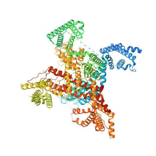
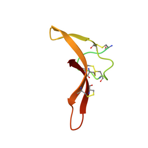
Cryo-EM reveals an unprecedented binding site for Na V 1.7 inhibitors enabling rational design of potent hybrid inhibitors.

Kschonsak, M.Jao, C.C.Arthur, C.P.Rohou, A.L.Bergeron, P.Ortwine, D.F.McKerrall, S.J.Hackos, D.H.Deng, L.Chen, J.Li, T.Dragovich, P.S.Volgraf, M.Wright, M.R.Payandeh, J.Ciferri, C.Tellis, J.C.

(2023) Elife 12

  • DOI: https://doi.org/10.7554/eLife.84151
  • Primary Citation of Related Structures:  
    8F0P, 8F0Q, 8F0R, 8F0S

  • PubMed Abstract: 

    The voltage-gated sodium (Na V ) channel Na V 1.7 has been identified as a potential novel analgesic target due to its involvement in human pain syndromes. However, clinically available Na V channel-blocking drugs are not selective among the nine Na V channel subtypes, Na V 1.1-Na V 1.9. Moreover, the two currently known classes of Na V 1.7 subtype-selective inhibitors (aryl- and acylsulfonamides) have undesirable characteristics that may limit their development. To this point understanding of the structure-activity relationships of the acylsulfonamide class of Na V 1.7 inhibitors, exemplified by the clinical development candidate GDC-0310 , has been based solely on a single co-crystal structure of an arylsulfonamide inhibitor bound to voltage-sensing domain 4 (VSD4). To advance inhibitor design targeting the Na V 1.7 channel, we pursued high-resolution ligand-bound Na V 1.7-VSD4 structures using cryogenic electron microscopy (cryo-EM). Here, we report that GDC-0310 engages the Na V 1.7-VSD4 through an unexpected binding mode orthogonal to the arylsulfonamide inhibitor class binding pose, which identifies a previously unknown ligand binding site in Na V channels. This finding enabled the design of a novel hybrid inhibitor series that bridges the aryl- and acylsulfonamide binding pockets and allows for the generation of molecules with substantially differentiated structures and properties. Overall, our study highlights the power of cryo-EM methods to pursue challenging drug targets using iterative and high-resolution structure-guided inhibitor design. This work also underscores an important role of the membrane bilayer in the optimization of selective Na V channel modulators targeting VSD4.


  • Organizational Affiliation

    Genentech Inc, Structural Biology, South San Francisco, United States.


Macromolecules
Find similar proteins by:  (by identity cutoff)  |  3D Structure
Entity ID: 1
MoleculeChains Sequence LengthOrganismDetailsImage
Sodium channel protein PaFPC1,Sodium channel protein type 9 subunit alpha chimera1,608Homo sapiensPeriplaneta americanaMutation(s): 0 
Membrane Entity: Yes 
UniProt & NIH Common Fund Data Resources
Find proteins for Q15858 (Homo sapiens)
Explore Q15858 
Go to UniProtKB:  Q15858
PHAROS:  Q15858
GTEx:  ENSG00000169432 
Find proteins for D0E0C2 (Periplaneta americana)
Explore D0E0C2 
Go to UniProtKB:  D0E0C2
Entity Groups  
Sequence Clusters30% Identity50% Identity70% Identity90% Identity95% Identity100% Identity
UniProt GroupsD0E0C2Q15858
Sequence Annotations
Expand
  • Reference Sequence
Find similar proteins by:  (by identity cutoff)  |  3D Structure
Entity ID: 2
MoleculeChains Sequence LengthOrganismDetailsImage
Beta-diguetoxin-Dc1a71Diguetia canitiesMutation(s): 0 
Membrane Entity: Yes 
UniProt
Find proteins for P49126 (Diguetia canities)
Explore P49126 
Go to UniProtKB:  P49126
Entity Groups  
Sequence Clusters30% Identity50% Identity70% Identity90% Identity95% Identity100% Identity
UniProt GroupP49126
Sequence Annotations
Expand
  • Reference Sequence
Oligosaccharides

Help

Entity ID: 3
MoleculeChains Length2D Diagram Glycosylation3D Interactions
beta-D-mannopyranose-(1-3)-[beta-D-mannopyranose-(1-6)]beta-D-mannopyranose-(1-3)-2-acetamido-2-deoxy-beta-D-glucopyranose-(1-3)-2-acetamido-2-deoxy-beta-D-glucopyranose
C
5N/AN/A
Glycosylation Resources
GlyTouCan:  G61217QH
GlyCosmos:  G61217QH
GlyGen:  G61217QH
Entity ID: 4
MoleculeChains Length2D Diagram Glycosylation3D Interactions
2-acetamido-2-deoxy-beta-D-glucopyranose-(1-4)-2-acetamido-2-deoxy-beta-D-glucopyranose
D
2N/AN/A
Glycosylation Resources
GlyTouCan:  G42666HT
GlyCosmos:  G42666HT
GlyGen:  G42666HT
Small Molecules
Ligands 4 Unique
IDChains Name / Formula / InChI Key2D Diagram3D Interactions
PEE
Query on PEE

Download Ideal Coordinates CCD File 
J [auth A]1,2-dioleoyl-sn-glycero-3-phosphoethanolamine
C41 H78 N O8 P
MWRBNPKJOOWZPW-NYVOMTAGSA-N
X80 (Subject of Investigation/LOI)
Query on X80

Download Ideal Coordinates CCD File 
I [auth A]5-chloro-4-(cyclopentylmethoxy)-N-(4-{[(1S,2S)-2-(dimethylamino)cyclohexyl]amino}-2-fluorobenzene-1-sulfonyl)-2-fluorobenzamide
C27 H34 Cl F2 N3 O4 S
ONUSAXHNRAIZSE-ZEQRLZLVSA-N
Y01
Query on Y01

Download Ideal Coordinates CCD File 
K [auth A]CHOLESTEROL HEMISUCCINATE
C31 H50 O4
WLNARFZDISHUGS-MIXBDBMTSA-N
NAG
Query on NAG

Download Ideal Coordinates CCD File 
E [auth A],
F [auth A],
G [auth A],
H [auth A]
2-acetamido-2-deoxy-beta-D-glucopyranose
C8 H15 N O6
OVRNDRQMDRJTHS-FMDGEEDCSA-N
Experimental Data & Validation

Experimental Data

  • Method: ELECTRON MICROSCOPY
  • Resolution: 3.10 Å
  • Aggregation State: PARTICLE 
  • Reconstruction Method: SINGLE PARTICLE 
EM Software:
TaskSoftware PackageVersion
MODEL REFINEMENTPHENIX1.20
RECONSTRUCTIONPHENIX1.20

Structure Validation

View Full Validation Report


Entry History & Funding Information

Deposition Data


Funding OrganizationLocationGrant Number
Not funded--

Revision History  (Full details and data files)

  • Version 1.0: 2023-04-12
    Type: Initial release