8E4R

Room-temperature X-ray structure of SARS-CoV-2 main protease H41A miniprecursor mutant in complex with GC373


Experimental Data Snapshot

  • Method: X-RAY DIFFRACTION
  • Resolution: 1.80 Å
  • R-Value Free: 0.195 
  • R-Value Work: 0.165 
  • R-Value Observed: 0.166 

wwPDB Validation   3D Report Full Report


Ligand Structure Quality Assessment 


This is version 1.2 of the entry. See complete history


Literature

Unmasking the Conformational Stability and Inhibitor Binding to SARS-CoV-2 Main Protease Active Site Mutants and Miniprecursor.

Kovalevsky, A.Coates, L.Kneller, D.W.Ghirlando, R.Aniana, A.Nashed, N.T.Louis, J.M.

(2022) J Mol Biol 434: 167876-167876

  • DOI: https://doi.org/10.1016/j.jmb.2022.167876
  • Primary Citation of Related Structures:  
    8E4J, 8E4R

  • PubMed Abstract: 

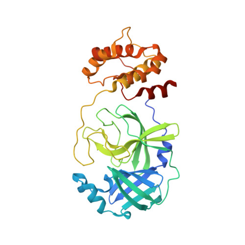
    We recently demonstrated that inhibitor binding reorganizes the oxyanion loop of a monomeric catalytic domain of SARS CoV-2 main protease (MPro) from an unwound (E) to a wound (active, E*) conformation, independent of dimerization. Here we assess the effect of the flanking N-terminal residues, to imitate the MPro precursor prior to its autoprocessing, on conformational equilibria rendering stability and inhibitor binding. Thermal denaturation (T m ) of C145A mutant, unlike H41A, increases by 6.8 °C, relative to wild-type mature dimer. An inactivating H41A mutation to maintain a miniprecursor containing TSAVL[Q or E] of the flanking nsp4 sequence in an intact form [ (-6) MPro H41A and (-6*) MPro H41A , respectively], and its corresponding mature MPro H41A were systematically examined. While the H41A mutation exerts negligible effect on T m and dimer dissociation constant (K dimer ) of MPro H41A , relative to the wild type MPro, both miniprecursors show a 4-5 °C decrease in T m and > 85-fold increase in K dimer as compared to MPro H41A . The K d for the binding of the covalent inhibitor GC373 to (-6*) MPro H41A increases ∼12-fold, relative to MPro H41A , concomitant with its dimerization. While the inhibitor-free dimer exhibits a state in transit from E to E* with a conformational asymmetry of the protomers' oxyanion loops and helical domains, inhibitor binding restores the asymmetry to mature-like oxyanion loop conformations (E*) but not of the helical domains. Disorder of the terminal residues 1-2 and 302-306 observed in both structures suggest that N-terminal autoprocessing is tightly coupled to the E-E* equilibrium and stable dimer formation.


  • Organizational Affiliation

    Neutron Scattering Division, Oak Ridge National Laboratory, 1 Bethel Valley Road, Oak Ridge, TN 37831, USA. Electronic address: kovalevskyay@ornl.gov.


Macromolecules
Find similar proteins by:  (by identity cutoff)  |  3D Structure
Entity ID: 1
MoleculeChains Sequence LengthOrganismDetailsImage
Replicase polyprotein 1ab
A, B
314Severe acute respiratory syndrome coronavirus 2Mutation(s): 2 
Gene Names: rep1a-1b
UniProt
Find proteins for P0DTD1 (Severe acute respiratory syndrome coronavirus 2)
Explore P0DTD1 
Go to UniProtKB:  P0DTD1
Entity Groups  
Sequence Clusters30% Identity50% Identity70% Identity90% Identity95% Identity100% Identity
UniProt GroupP0DTD1
Sequence Annotations
Expand
  • Reference Sequence
Small Molecules
Ligands 1 Unique
IDChains Name / Formula / InChI Key2D Diagram3D Interactions
K36 (Subject of Investigation/LOI)
Query on K36

Download Ideal Coordinates CCD File 
C [auth A],
D [auth B]
(1S,2S)-2-({N-[(benzyloxy)carbonyl]-L-leucyl}amino)-1-hydroxy-3-[(3S)-2-oxopyrrolidin-3-yl]propane-1-sulfonic acid
C21 H31 N3 O8 S
BSPZFJDYQHDZNR-HTCLRFROSA-N
Biologically Interesting Molecules (External Reference) 1 Unique
Experimental Data & Validation

Experimental Data

  • Method: X-RAY DIFFRACTION
  • Resolution: 1.80 Å
  • R-Value Free: 0.195 
  • R-Value Work: 0.165 
  • R-Value Observed: 0.166 
  • Space Group: P 1 21 1
Unit Cell:
Length ( Å )Angle ( ˚ )
a = 55.843α = 90
b = 100.126β = 108.34
c = 59.957γ = 90
Software Package:
Software NamePurpose
PHENIXrefinement
CrysalisProdata reduction
CrysalisProdata scaling
PHASERphasing

Structure Validation

View Full Validation Report


Ligand Structure Quality Assessment 


Entry History & Funding Information

Deposition Data


Funding OrganizationLocationGrant Number
Not funded--

Revision History  (Full details and data files)

  • Version 1.0: 2022-11-23
    Type: Initial release
  • Version 1.1: 2022-11-30
    Changes: Database references
  • Version 1.2: 2023-10-25
    Changes: Data collection, Refinement description