8DYA

Structure of the SARS-CoV-2 spike glycoprotein S2 subunit


Experimental Data Snapshot

  • Method: ELECTRON MICROSCOPY
  • Resolution: 3.67 Å
  • Aggregation State: PARTICLE 
  • Reconstruction Method: SINGLE PARTICLE 

wwPDB Validation   3D Report Full Report


This is version 1.2 of the entry. See complete history


Literature

SARS-CoV-2 spike conformation determines plasma neutralizing activity elicited by a wide panel of human vaccines.

Bowen, J.E.Park, Y.J.Stewart, C.Brown, J.T.Sharkey, W.K.Walls, A.C.Joshi, A.Sprouse, K.R.McCallum, M.Tortorici, M.A.Franko, N.M.Logue, J.K.Mazzitelli, I.G.Nguyen, A.W.Silva, R.P.Huang, Y.Low, J.S.Jerak, J.Tiles, S.W.Ahmed, K.Shariq, A.Dan, J.M.Zhang, Z.Weiskopf, D.Sette, A.Snell, G.Posavad, C.M.Iqbal, N.T.Geffner, J.Bandera, A.Gori, A.Sallusto, F.Maynard, J.A.Crotty, S.Van Voorhis, W.C.Simmerling, C.Grifantini, R.Chu, H.Y.Corti, D.Veesler, D.

(2022) Sci Immunol 7: eadf1421-eadf1421

  • DOI: https://doi.org/10.1126/sciimmunol.adf1421
  • Primary Citation of Related Structures:  
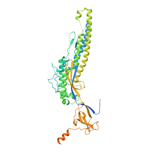
    8DYA

  • PubMed Abstract: 

    Numerous safe and effective coronavirus disease 2019 vaccines have been developed worldwide that use various delivery technologies and engineering strategies. We show here that vaccines containing prefusion-stabilizing S mutations elicit antibody responses in humans with enhanced recognition of S and the S 1 subunit relative to postfusion S as compared with vaccines lacking these mutations or natural infection. Prefusion S and S 1 antibody binding titers positively and equivalently correlated with neutralizing activity, and depletion of S 1 -directed antibodies completely abrogated plasma neutralizing activity. We show that neutralizing activity is almost entirely directed to the S 1 subunit and that variant cross-neutralization is mediated solely by receptor binding domain-specific antibodies. Our data provide a quantitative framework for guiding future S engineering efforts to develop vaccines with higher resilience to the emergence of variants than current technologies.


  • Organizational Affiliation

    Department of Biochemistry, University of Washington, Seattle, WA 98195, USA.


Macromolecules
Find similar proteins by:  (by identity cutoff)  |  3D Structure
Entity ID: 1
MoleculeChains Sequence LengthOrganismDetailsImage
Spike glycoprotein
A, B, C
624Severe acute respiratory syndrome coronavirus 2Mutation(s): 8 
UniProt
Find proteins for P0DTC2 (Severe acute respiratory syndrome coronavirus 2)
Explore P0DTC2 
Go to UniProtKB:  P0DTC2
Entity Groups  
Sequence Clusters30% Identity50% Identity70% Identity90% Identity95% Identity100% Identity
UniProt GroupP0DTC2
Sequence Annotations
Expand
  • Reference Sequence
Small Molecules
Ligands 1 Unique
IDChains Name / Formula / InChI Key2D Diagram3D Interactions
NAG
Query on NAG

Download Ideal Coordinates CCD File 
D [auth A]
E [auth A]
F [auth A]
G [auth A]
H [auth A]
D [auth A],
E [auth A],
F [auth A],
G [auth A],
H [auth A],
I [auth A],
J [auth B],
K [auth B],
L [auth B],
M [auth B],
N [auth B],
O [auth B],
P [auth C],
Q [auth C],
R [auth C],
S [auth C],
T [auth C],
U [auth C]
2-acetamido-2-deoxy-beta-D-glucopyranose
C8 H15 N O6
OVRNDRQMDRJTHS-FMDGEEDCSA-N
Experimental Data & Validation

Experimental Data

  • Method: ELECTRON MICROSCOPY
  • Resolution: 3.67 Å
  • Aggregation State: PARTICLE 
  • Reconstruction Method: SINGLE PARTICLE 
EM Software:
TaskSoftware PackageVersion
RECONSTRUCTIONcryoSPARC

Structure Validation

View Full Validation Report


Entry History & Funding Information

Deposition Data


Funding OrganizationLocationGrant Number
National Institutes of Health/National Institute Of Allergy and Infectious Diseases (NIH/NIAID)United States--

Revision History  (Full details and data files)

  • Version 1.0: 2022-11-30
    Type: Initial release
  • Version 1.1: 2023-01-04
    Changes: Database references
  • Version 1.2: 2023-01-11
    Changes: Database references