8DSU

Crystal Structure of SARS CoV-2 Mpro with Pfizer Intravenous Inhibitor PF-00835231


Experimental Data Snapshot

  • Method: X-RAY DIFFRACTION
  • Resolution: 1.86 Å
  • R-Value Free: 0.254 
  • R-Value Work: 0.205 
  • R-Value Observed: 0.207 

wwPDB Validation   3D Report Full Report


Ligand Structure Quality Assessment 


This is version 1.3 of the entry. See complete history


Literature

Crystal Structures of Inhibitor-Bound Main Protease from Delta- and Gamma-Coronaviruses.

Zvornicanin, S.N.Shaqra, A.M.Huang, Q.J.Ornelas, E.Moghe, M.Knapp, M.Moquin, S.Dovala, D.Schiffer, C.A.Kurt Yilmaz, N.

(2023) Viruses 15

  • DOI: https://doi.org/10.3390/v15030781
  • Primary Citation of Related Structures:  
    8DSU, 8E7C, 8E7N, 8FWX

  • PubMed Abstract: 

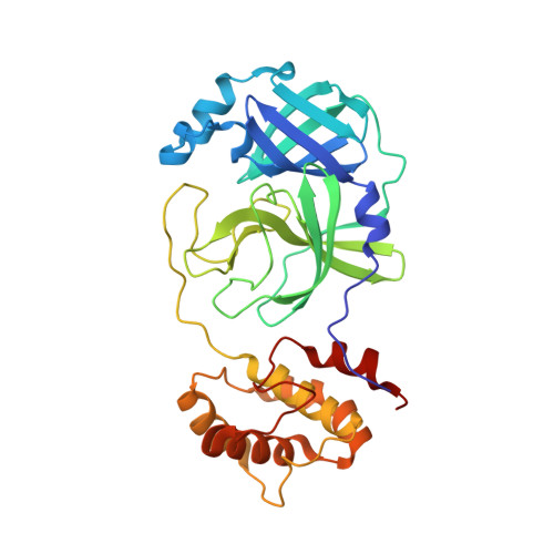
    With the spread of SARS-CoV-2 throughout the globe causing the COVID-19 pandemic, the threat of zoonotic transmissions of coronaviruses (CoV) has become even more evident. As human infections have been caused by alpha- and beta-CoVs, structural characterization and inhibitor design mostly focused on these two genera. However, viruses from the delta and gamma genera also infect mammals and pose a potential zoonotic transmission threat. Here, we determined the inhibitor-bound crystal structures of the main protease (M pro ) from the delta-CoV porcine HKU15 and gamma-CoV SW1 from the beluga whale. A comparison with the apo structure of SW1 M pro , which is also presented here, enabled the identification of structural arrangements upon inhibitor binding at the active site. The cocrystal structures reveal binding modes and interactions of two covalent inhibitors, PF-00835231 (active form of lufotrelvir) bound to HKU15, and GC376 bound to SW1 M pro . These structures may be leveraged to target diverse coronaviruses and toward the structure-based design of pan-CoV inhibitors.


  • Organizational Affiliation

    Department of Biochemistry and Molecular Biotechnology, University of Massachusetts Chan Medical School, Worcester, MA 01605, USA.


Macromolecules
Find similar proteins by:  (by identity cutoff)  |  3D Structure
Entity ID: 1
MoleculeChains Sequence LengthOrganismDetailsImage
3C-like proteinase nsp5
A, B
306Severe acute respiratory syndrome coronavirus 2Mutation(s): 0 
Gene Names: rep1a-1b
EC: 3.4.22.69
UniProt
Find proteins for P0DTD1 (Severe acute respiratory syndrome coronavirus 2)
Explore P0DTD1 
Go to UniProtKB:  P0DTD1
Entity Groups  
Sequence Clusters30% Identity50% Identity70% Identity90% Identity95% Identity100% Identity
UniProt GroupP0DTD1
Sequence Annotations
Expand
  • Reference Sequence
Experimental Data & Validation

Experimental Data

  • Method: X-RAY DIFFRACTION
  • Resolution: 1.86 Å
  • R-Value Free: 0.254 
  • R-Value Work: 0.205 
  • R-Value Observed: 0.207 
  • Space Group: P 1 21 1
Unit Cell:
Length ( Å )Angle ( ˚ )
a = 54.896α = 90
b = 98.937β = 107.624
c = 59.148γ = 90
Software Package:
Software NamePurpose
PHENIXrefinement
PHENIXrefinement
Cootmodel building
PHASERphasing
CrysalisProdata reduction
CrysalisProdata scaling
CrysalisProdata collection

Structure Validation

View Full Validation Report


Ligand Structure Quality Assessment 


Entry History & Funding Information

Deposition Data


Funding OrganizationLocationGrant Number
Other privateUnited States--

Revision History  (Full details and data files)

  • Version 1.0: 2023-03-29
    Type: Initial release
  • Version 1.1: 2023-04-05
    Changes: Derived calculations
  • Version 1.2: 2023-04-12
    Changes: Database references
  • Version 1.3: 2023-10-25
    Changes: Data collection, Refinement description