8DPM

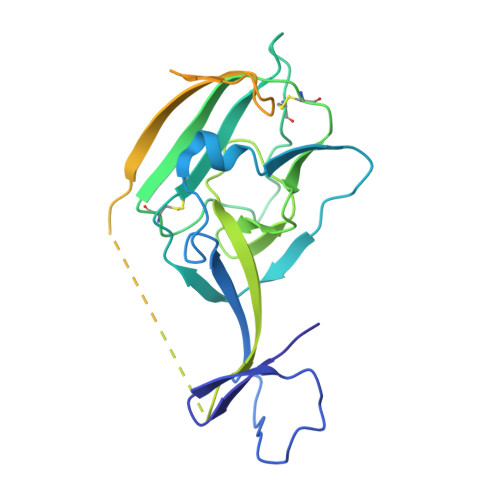
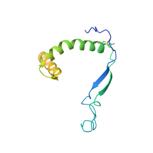
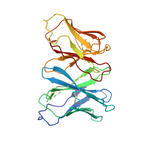
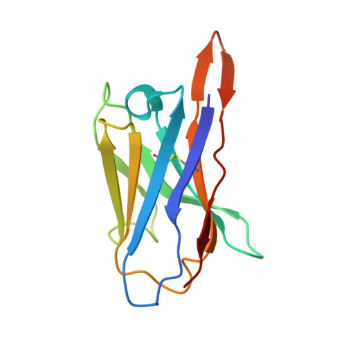
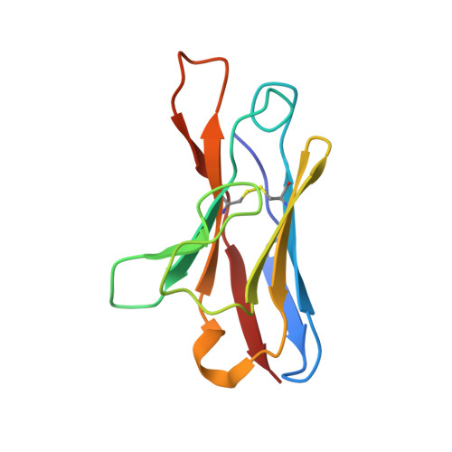
Structure of EBOV GP lacking the mucin-like domain with 9.20.1A2 Fab and 6D6 scFv bound


Experimental Data Snapshot

  • Method: ELECTRON MICROSCOPY
  • Resolution: 3.00 Å
  • Aggregation State: PARTICLE 
  • Reconstruction Method: SINGLE PARTICLE 

wwPDB Validation   3D Report Full Report


This is version 1.1 of the entry. See complete history


Literature

The evolution and determinants of neutralization of potent head-binding antibodies against Ebola virus.

Yu, X.Hastie, K.M.Davis, C.W.Avalos, R.D.Williams, D.Parekh, D.Hui, S.Mann, C.Hariharan, C.Takada, A.Ahmed, R.Saphire, E.O.

(2023) Cell Rep 42: 113366-113366

  • DOI: https://doi.org/10.1016/j.celrep.2023.113366
  • Primary Citation of Related Structures:  
    8DPL, 8DPM

  • PubMed Abstract: 

    Monoclonal antibodies against the Ebola virus (EBOV) surface glycoprotein are effective treatments for EBOV disease. Antibodies targeting the EBOV glycoprotein (GP) head epitope have potent neutralization and Fc effector function activity and thus are of high interest as therapeutics and for vaccine design. Here we focus on the head-binding antibodies 1A2 and 1D5, which have been identified previously in a longitudinal study of survivors of EBOV infection. 1A2 and 1D5 have the same heavy- and light-chain germlines despite being isolated from different individuals and at different time points after recovery from infection. Cryoelectron microscopy analysis of each antibody in complex with the EBOV surface GP reveals key amino acid substitutions in 1A2 that contribute to greater affinity, improved neutralization potency, and enhanced breadth as well as two strategies for antibody evolution from a common site.


  • Organizational Affiliation

    Center for Infectious Disease and Vaccine Discovery, La Jolla Institute for Immunology, La Jolla, CA 92037, USA.


Macromolecules
Find similar proteins by:  (by identity cutoff)  |  3D Structure
Entity ID: 1
MoleculeChains Sequence LengthOrganismDetailsImage
Glycoprotein GP1A [auth F],
F [auth A],
K
280Ebola virus - Mayinga, Zaire, 1976Mutation(s): 0 
Gene Names: GP
UniProt
Find proteins for Q05320 (Zaire ebolavirus (strain Mayinga-76))
Explore Q05320 
Go to UniProtKB:  Q05320
Entity Groups  
Sequence Clusters30% Identity50% Identity70% Identity90% Identity95% Identity100% Identity
UniProt GroupQ05320
Sequence Annotations
Expand
  • Reference Sequence
Find similar proteins by:  (by identity cutoff)  |  3D Structure
Entity ID: 2
MoleculeChains Sequence LengthOrganismDetailsImage
Glycoprotein GP2B [auth G],
G [auth B],
L
136Ebola virus - Mayinga, Zaire, 1976Mutation(s): 0 
Gene Names: GP
UniProt
Find proteins for Q05320 (Zaire ebolavirus (strain Mayinga-76))
Explore Q05320 
Go to UniProtKB:  Q05320
Entity Groups  
Sequence Clusters30% Identity50% Identity70% Identity90% Identity95% Identity100% Identity
UniProt GroupQ05320
Sequence Annotations
Expand
  • Reference Sequence
Find similar proteins by:  (by identity cutoff)  |  3D Structure
Entity ID: 3
MoleculeChains Sequence LengthOrganismDetailsImage
Antibody 6D6 scFvC [auth H],
H [auth C],
M
243Mus musculusMutation(s): 0 
Entity Groups  
Sequence Clusters30% Identity50% Identity70% Identity90% Identity95% Identity100% Identity
Sequence Annotations
Expand
  • Reference Sequence
Find similar proteins by:  (by identity cutoff)  |  3D Structure
Entity ID: 4
MoleculeChains Sequence LengthOrganismDetailsImage
Antibody 9.20.1A2 Fab heavy chainD [auth I],
I [auth D],
N
122Homo sapiensMutation(s): 0 
Entity Groups  
Sequence Clusters30% Identity50% Identity70% Identity90% Identity95% Identity100% Identity
Sequence Annotations
Expand
  • Reference Sequence
Find similar proteins by:  (by identity cutoff)  |  3D Structure
Entity ID: 5
MoleculeChains Sequence LengthOrganismDetailsImage
Antibody 9.20.1A2 Fab light chainE [auth J],
J [auth E],
O
112Homo sapiensMutation(s): 0 
Entity Groups  
Sequence Clusters30% Identity50% Identity70% Identity90% Identity95% Identity100% Identity
Sequence Annotations
Expand
  • Reference Sequence
Oligosaccharides

Help

Entity ID: 6
MoleculeChains Length2D Diagram Glycosylation3D Interactions
alpha-D-mannopyranose-(1-3)-alpha-D-mannopyranose-(1-6)-[alpha-D-mannopyranose-(1-3)]beta-D-mannopyranose-(1-4)-2-acetamido-2-deoxy-beta-D-glucopyranose-(1-4)-2-acetamido-2-deoxy-beta-D-glucopyranose
P, Q, R
6N-Glycosylation
Glycosylation Resources
GlyTouCan:  G09724ZC
GlyCosmos:  G09724ZC
GlyGen:  G09724ZC
Experimental Data & Validation

Experimental Data

  • Method: ELECTRON MICROSCOPY
  • Resolution: 3.00 Å
  • Aggregation State: PARTICLE 
  • Reconstruction Method: SINGLE PARTICLE 

Structure Validation

View Full Validation Report


Entry History & Funding Information

Deposition Data


Funding OrganizationLocationGrant Number
National Institutes of Health/National Institute Of Allergy and Infectious Diseases (NIH/NIAID)United StatesU19 AI142790
National Science Foundation (NSF, United States)United StatesNSF 1531991

Revision History  (Full details and data files)

  • Version 1.0: 2023-07-19
    Type: Initial release
  • Version 1.1: 2023-11-22
    Changes: Data collection, Database references