


Crystal Structure of SARS CoV-2 NSP15 Endroribonuclease H250A
Farraj, R.A., Edwards, R.A., Glover, J.N.M.To be published.
Experimental Data Snapshot
wwPDB Validation 3D Report Full Report

Entity ID: 1 | |||||
|---|---|---|---|---|---|
| Molecule | Chains | Sequence Length | Organism | Details | Image |
| Uridylate-specific endoribonuclease nsp15 | 373 | Severe acute respiratory syndrome coronavirus 2 | Mutation(s): 1 Gene Names: rep, 1a-1b EC: 4.6.1 | 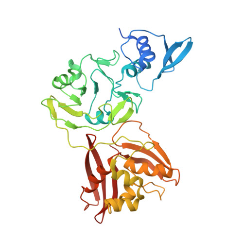 | |
UniProt | |||||
Find proteins for P0DTD1 (Severe acute respiratory syndrome coronavirus 2) Explore P0DTD1 Go to UniProtKB: P0DTD1 | |||||
Entity Groups | |||||
| Sequence Clusters | 30% Identity50% Identity70% Identity90% Identity95% Identity100% Identity | ||||
| UniProt Group | P0DTD1 | ||||
Sequence AnnotationsExpand | |||||
| |||||
| Length ( Å ) | Angle ( ˚ ) |
|---|---|
| a = 152.525 | α = 90 |
| b = 152.525 | β = 90 |
| c = 109.644 | γ = 120 |
| Software Name | Purpose |
|---|---|
| PHENIX | refinement |
| HKL-2000 | data reduction |
| SCALEPACK | data scaling |
| PHASER | phasing |
| Funding Organization | Location | Grant Number |
|---|---|---|
| Canadian Institutes of Health Research (CIHR) | Canada | -- |
RCSB PDB (citation) is hosted by
RCSB PDB is a member of the

