8CSE

WbbB in complex with alpha-Rha-(1-3)-beta-GlcNAc acceptor


Experimental Data Snapshot

  • Method: X-RAY DIFFRACTION
  • Resolution: 2.30 Å
  • R-Value Free: 0.225 
  • R-Value Work: 0.167 
  • R-Value Observed: 0.170 

wwPDB Validation   3D Report Full Report


Ligand Structure Quality Assessment 


This is version 2.0 of the entry. See complete history


Literature

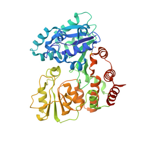
The retaining beta-Kdo glycosyltransferase WbbB uses a double-displacement mechanism with an intermediate adduct rearrangement step.

Forrester, T.J.B.Ovchinnikova, O.G.Li, Z.Kitova, E.N.Nothof, J.T.Koizumi, A.Klassen, J.S.Lowary, T.L.Whitfield, C.Kimber, M.S.

(2022) Nat Commun 13: 6277-6277

  • DOI: https://doi.org/10.1038/s41467-022-33988-1
  • Primary Citation of Related Structures:  
    8CSB, 8CSC, 8CSD, 8CSE, 8CSF

  • PubMed Abstract: 

    WbbB, a lipopolysaccharide O-antigen synthesis enzyme from Raoultella terrigena, contains an N-terminal glycosyltransferase domain with a highly modified architecture that adds a terminal β-Kdo (3-deoxy-D-manno-oct-2-ulosonic acid) residue to the O-antigen saccharide, with retention of stereochemistry. We show, using mass spectrometry, that WbbB forms a covalent adduct between the catalytic nucleophile, Asp232, and Kdo. We also determine X-ray structures for the CMP-β-Kdo donor complex, for Kdo-adducts with D232N and D232C WbbB variants, for a synthetic disaccharide acceptor complex, and for a ternary complex with both a Kdo-adduct and the acceptor. Together, these structures show that the enzyme-linked Asp232-Kdo adduct rotates to reposition the Kdo into a second sub-site, which then transfers Kdo to the acceptor. Retaining glycosyltransferases were thought to use only the front-side S N i substitution mechanism; here we show that retaining glycosyltransferases can also potentially use double-displacement mechanisms, but incorporating an additional catalytic subsite requires rearrangement of the protein's architecture.


  • Organizational Affiliation

    Department of Molecular and Cellular Biology, University of Guelph, 50 Stone Road E., Guelph, ON, N1G 2W1, Canada.


Macromolecules
Find similar proteins by:  (by identity cutoff)  |  3D Structure
Entity ID: 1
MoleculeChains Sequence LengthOrganismDetailsImage
N-acetyl glucosaminyl transferase
A, B
410Raoultella terrigenaMutation(s): 0 
Gene Names: wbbB
UniProt
Find proteins for Q6U8B0 (Raoultella terrigena)
Explore Q6U8B0 
Go to UniProtKB:  Q6U8B0
Entity Groups  
Sequence Clusters30% Identity50% Identity70% Identity90% Identity95% Identity100% Identity
UniProt GroupQ6U8B0
Sequence Annotations
Expand
  • Reference Sequence
Oligosaccharides

Help

Entity ID: 2
MoleculeChains Length2D Diagram Glycosylation3D Interactions
alpha-L-rhamnopyranose-(1-3)-2-acetamido-2-deoxy-beta-D-glucopyranoseC [auth D],
D [auth E],
E [auth F],
F [auth G],
G [auth H]
2N/A
Glycosylation Resources
GlyTouCan:  G99436AO
GlyCosmos:  G99436AO
Small Molecules
Modified Residues  1 Unique
IDChains TypeFormula2D DiagramParent
CSO
Query on CSO
A, B
L-PEPTIDE LINKINGC3 H7 N O3 SCYS
Experimental Data & Validation

Experimental Data

  • Method: X-RAY DIFFRACTION
  • Resolution: 2.30 Å
  • R-Value Free: 0.225 
  • R-Value Work: 0.167 
  • R-Value Observed: 0.170 
  • Space Group: C 2 2 21
Unit Cell:
Length ( Å )Angle ( ˚ )
a = 92.38α = 90
b = 158.02β = 90
c = 117.58γ = 90
Software Package:
Software NamePurpose
PHENIXrefinement
XDSdata reduction
XSCALEdata scaling
PHASERphasing

Structure Validation

View Full Validation Report


Ligand Structure Quality Assessment 


Entry History & Funding Information

Deposition Data


Funding OrganizationLocationGrant Number
Natural Sciences and Engineering Research Council (NSERC, Canada)Canada04045-2015

Revision History  (Full details and data files)

  • Version 1.0: 2022-11-09
    Type: Initial release
  • Version 1.1: 2023-10-18
    Changes: Data collection, Refinement description
  • Version 2.0: 2023-11-15
    Changes: Advisory, Atomic model, Data collection, Database references