


Structure of West Nile Virus NS2B-NS3 protease - to be published
Fairhead, M., Godoy, A.S., Balcomb, B.H., von Delft, F.To be published.
Experimental Data Snapshot
wwPDB Validation 3D Report Full Report

Entity ID: 1 | |||||
|---|---|---|---|---|---|
| Molecule | Chains | Sequence Length | Organism | Details | Image |
| Serine protease subunit NS2B,Serine protease/Helicase NS3 | 245 | West Nile virus | Mutation(s): 1 Gene Names: GP1, MZ11_60484gpGP1, MZ11_60553gpGP1 EC: 3.4.21.91 (PDB Primary Data), 3.6.1.15 (PDB Primary Data), 3.6.4.13 (PDB Primary Data) | 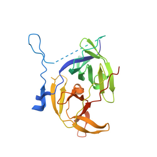 | |
UniProt | |||||
Find proteins for Q9Q6P4 (West Nile virus (strain NY-99)) Explore Q9Q6P4 Go to UniProtKB: Q9Q6P4 | |||||
Entity Groups | |||||
| Sequence Clusters | 30% Identity50% Identity70% Identity90% Identity95% Identity100% Identity | ||||
| UniProt Group | Q9Q6P4 | ||||
Sequence AnnotationsExpand | |||||
| |||||
| Length ( Å ) | Angle ( ˚ ) |
|---|---|
| a = 56.895 | α = 90 |
| b = 56.895 | β = 90 |
| c = 103.902 | γ = 120 |
| Software Name | Purpose |
|---|---|
| PHENIX | refinement |
| xia2 | data reduction |
| xia2 | data scaling |
| PHASER | phasing |
| Funding Organization | Location | Grant Number |
|---|---|---|
| National Institutes of Health/National Institute Of Allergy and Infectious Diseases (NIH/NIAID) | United States | U19AI171399 |
RCSB PDB (citation) is hosted by
RCSB PDB is a member of the

