


Structure of EV D68 3C protease
Lithgo, R.M., von Delft, F.To be published.
Experimental Data Snapshot
wwPDB Validation 3D Report Full Report

Entity ID: 1 | |||||
|---|---|---|---|---|---|
| Molecule | Chains | Sequence Length | Organism | Details | Image |
| Protease 3C | 191 | Enterovirus | Mutation(s): 0 EC: 3.4.22.28 | 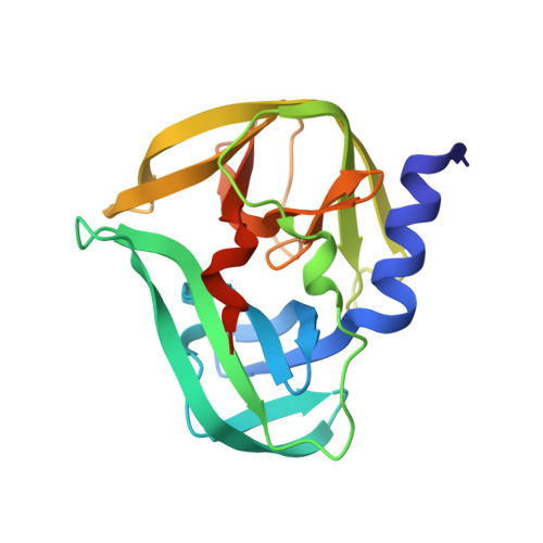 | |
UniProt | |||||
Find proteins for Q68T42 (Human enterovirus D68) Explore Q68T42 Go to UniProtKB: Q68T42 | |||||
Entity Groups | |||||
| Sequence Clusters | 30% Identity50% Identity70% Identity90% Identity95% Identity100% Identity | ||||
| UniProt Group | Q68T42 | ||||
Sequence AnnotationsExpand | |||||
| |||||
| Length ( Å ) | Angle ( ˚ ) |
|---|---|
| a = 42.82 | α = 90 |
| b = 62.53 | β = 90 |
| c = 147.36 | γ = 90 |
| Software Name | Purpose |
|---|---|
| REFMAC | refinement |
| xia2 | data reduction |
| xia2 | data scaling |
| PHASER | phasing |
| Funding Organization | Location | Grant Number |
|---|---|---|
| National Institutes of Health/National Institute Of Allergy and Infectious Diseases (NIH/NIAID) | United States | U19AI171399 |
RCSB PDB (citation) is hosted by
RCSB PDB is a member of the

