


Crystal structure of Paradendryphiella salina PL7C alginate lyase
Fredslund, F., Welner, D.W., Wilkens, C.To be published.
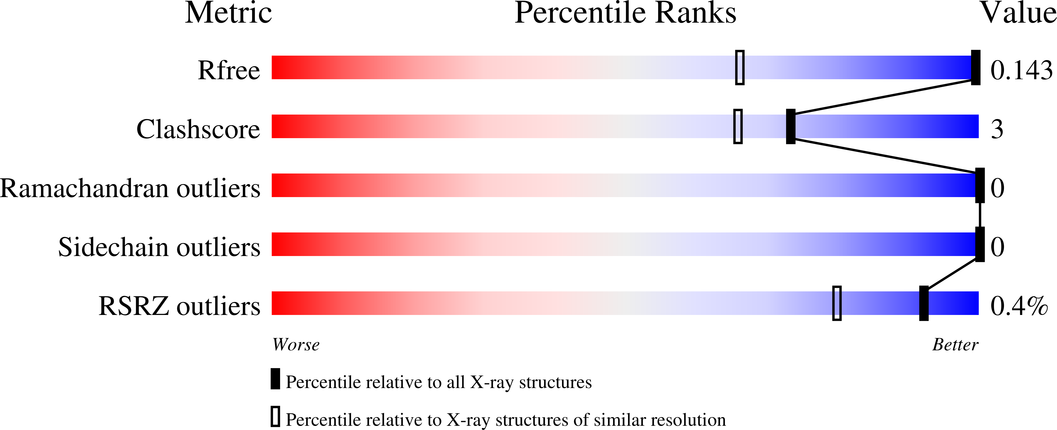
Experimental Data Snapshot
wwPDB Validation 3D Report Full Report
Entity ID: 1 | |||||
|---|---|---|---|---|---|
| Molecule | Chains | Sequence Length | Organism | Details | Image |
| Alginate lyase | 234 | Paradendryphiella salina | Mutation(s): 0 Gene Names: PsAlg7C | 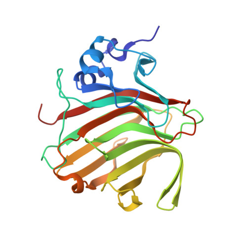 | |
UniProt | |||||
Find proteins for A0A7I9C8Z1 (Paradendryphiella salina) Explore A0A7I9C8Z1 Go to UniProtKB: A0A7I9C8Z1 | |||||
Entity Groups | |||||
| Sequence Clusters | 30% Identity50% Identity70% Identity90% Identity95% Identity100% Identity | ||||
| UniProt Group | A0A7I9C8Z1 | ||||
Sequence AnnotationsExpand | |||||
| |||||
| Ligands 1 Unique | |||||
|---|---|---|---|---|---|
| ID | Chains | Name / Formula / InChI Key | 2D Diagram | 3D Interactions | |
| EDO Query on EDO | B [auth A] | 1,2-ETHANEDIOL C2 H6 O2 LYCAIKOWRPUZTN-UHFFFAOYSA-N |  | ||
| Length ( Å ) | Angle ( ˚ ) |
|---|---|
| a = 41.21 | α = 90 |
| b = 59.8 | β = 90 |
| c = 90.07 | γ = 90 |
| Software Name | Purpose |
|---|---|
| MxCuBE | data collection |
| PHENIX | refinement |
| XDS | data reduction |
| XSCALE | data scaling |
| Coot | model building |
| PHASER | phasing |
| Funding Organization | Location | Grant Number |
|---|---|---|
| Other government | Denmark | -- |
RCSB PDB (citation) is hosted by
RCSB PDB is a member of the

