8C1N

FMDV 3D polymerase in complex with 3B1 protein solved in P212121 space group


Experimental Data Snapshot

  • Method: X-RAY DIFFRACTION
  • Resolution: 1.70 Å
  • R-Value Free: 0.238 
  • R-Value Work: 0.220 
  • R-Value Observed: 0.221 

wwPDB Validation   3D Report Full Report


Ligand Structure Quality Assessment 


This is version 1.1 of the entry. See complete history


Literature

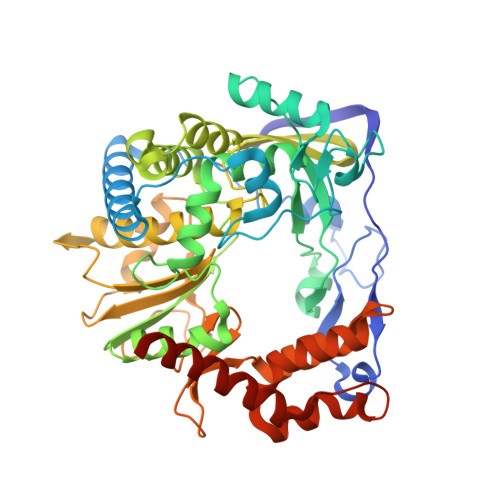
Dual role of the foot-and-mouth disease virus 3B1 protein in the replication complex: As protein primer and as an essential component to recruit 3Dpol to membranes.

Ferrer-Orta, C.Ferrero, D.S.Verdaguer, N.

(2023) PLoS Pathog 19: e1011373-e1011373

  • DOI: https://doi.org/10.1371/journal.ppat.1011373
  • Primary Citation of Related Structures:  
    8C1N, 8C2P

  • PubMed Abstract: 

    Picornavirus genome replication takes place in specialized intracellular membrane compartments that concentrate viral RNA and proteins as well as a number of host factors that also participate in the process. The core enzyme in the replication machinery is the viral RNA-dependent RNA polymerase (RdRP) 3Dpol. Replication requires the primer protein 3B (or VPg) attached to two uridine molecules. 3B uridylylation is also catalysed by 3Dpol. Another critical interaction in picornavirus replication is that between 3Dpol and the precursor 3AB, a membrane-binding protein responsible for the localization of 3Dpol to the membranous compartments at which replication occurs. Unlike other picornaviruses, the animal pathogen foot-and-mouth disease virus (FMDV), encodes three non-identical copies of the 3B (3B1, 3B2, and 3B3) that could be specialized in different functions within the replication complex. Here, we have used a combination of biophysics, molecular and structural biology approaches to characterize the functional binding of FMDV 3B1 to the base of the palm of 3Dpol. The 1.7 Å resolution crystal structure of the FMDV 3Dpol -3B1 complex shows that 3B1 simultaneously links two 3Dpol molecules by binding at the bottom of their palm subdomains in an almost symmetric way. The two 3B1 contact surfaces involve a combination of hydrophobic and basic residues at the N- (G5-P6, R9; Region I) and C-terminus (R16, L19-P20; Region II) of this small protein. Enzyme-Linked Immunosorbent Assays (ELISA) show that the two 3B1 binding sites play a role in 3Dpol binding, with region II presenting the highest affinity. ELISA assays show that 3Dpol has higher binding affinity for 3B1 than for 3B2 or 3B3. Membrane-based pull-down assays show that 3B1 region II, and to a lesser extent also region I play essential roles in mediating the interaction of 3AB with the polymerase and its recruitment to intracellular membranes.


  • Organizational Affiliation

    Instituto de Biología Molecular de Barcelona. Consejo Superior de Investigaciones Científicas (IBMB-CSIC), Barcelona, Spain.


Macromolecules
Find similar proteins by:  (by identity cutoff)  |  3D Structure
Entity ID: 1
MoleculeChains Sequence LengthOrganismDetailsImage
RNA-directed RNA polymerase 3D-POL
A, B
481Foot-and-mouth disease virusMutation(s): 0 
EC: 2.7.7.48
UniProt
Find proteins for P03311 (Foot-and-mouth disease virus (isolate -/Spain/S8c1SantaPau/1970 serotype C))
Explore P03311 
Go to UniProtKB:  P03311
Entity Groups  
Sequence Clusters30% Identity50% Identity70% Identity90% Identity95% Identity100% Identity
UniProt GroupP03311
Sequence Annotations
Expand
  • Reference Sequence

Find similar proteins by:  Sequence   |   3D Structure  

Entity ID: 2
MoleculeChains Sequence LengthOrganismDetailsImage
Protein 3B-123Foot-and-mouth disease virusMutation(s): 0 
UniProt
Find proteins for P03311 (Foot-and-mouth disease virus (isolate -/Spain/S8c1SantaPau/1970 serotype C))
Explore P03311 
Go to UniProtKB:  P03311
Entity Groups  
Sequence Clusters30% Identity50% Identity70% Identity90% Identity95% Identity100% Identity
UniProt GroupP03311
Sequence Annotations
Expand
  • Reference Sequence
Small Molecules
Ligands 3 Unique
IDChains Name / Formula / InChI Key2D Diagram3D Interactions
3PO (Subject of Investigation/LOI)
Query on 3PO

Download Ideal Coordinates CCD File 
D [auth A],
J [auth B]
TRIPHOSPHATE
H5 O10 P3
UNXRWKVEANCORM-UHFFFAOYSA-N
GOL
Query on GOL

Download Ideal Coordinates CCD File 
E [auth A]
F [auth A]
G [auth A]
H [auth A]
K [auth B]
E [auth A],
F [auth A],
G [auth A],
H [auth A],
K [auth B],
L [auth B],
M [auth B],
N [auth B],
O [auth B],
P [auth B],
Q [auth B]
GLYCEROL
C3 H8 O3
PEDCQBHIVMGVHV-UHFFFAOYSA-N
ACT
Query on ACT

Download Ideal Coordinates CCD File 
I [auth A],
R [auth B]
ACETATE ION
C2 H3 O2
QTBSBXVTEAMEQO-UHFFFAOYSA-M
Experimental Data & Validation

Experimental Data

  • Method: X-RAY DIFFRACTION
  • Resolution: 1.70 Å
  • R-Value Free: 0.238 
  • R-Value Work: 0.220 
  • R-Value Observed: 0.221 
  • Space Group: P 21 21 21
Unit Cell:
Length ( Å )Angle ( ˚ )
a = 93.343α = 90
b = 97.024β = 90
c = 115.902γ = 90
Software Package:
Software NamePurpose
PHENIXrefinement
DMphasing
SCALAdata scaling
autoPROCdata processing
XDSdata reduction

Structure Validation

View Full Validation Report


Ligand Structure Quality Assessment 


Entry History & Funding Information

Deposition Data


Funding OrganizationLocationGrant Number
Ministerio de Ciencia e Innovacion (MCIN)SpainPID2020-117976GB-100

Revision History  (Full details and data files)

  • Version 1.0: 2023-04-26
    Type: Initial release
  • Version 1.1: 2023-05-10
    Changes: Database references