Orthoparamyxovirinae C Proteins Have a Common Origin and a Common Structural Organization.
Roy, A., Chan Mine, E., Gaifas, L., Leyrat, C., Volchkova, V.A., Baudin, F., Martinez-Gil, L., Volchkov, V.E., Karlin, D.G., Bourhis, J.M., Jamin, M.(2023) Biomolecules 13
- PubMed: 36979390
- DOI: https://doi.org/10.3390/biom13030455
- Primary Citation of Related Structures:
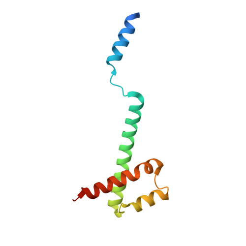
8BJW - PubMed Abstract:
The protein C is a small viral protein encoded in an overlapping frame of the P gene in the subfamily Orthoparamyxovirinae. This protein, expressed by alternative translation initiation, is a virulence factor that regulates viral transcription, replication, and production of defective interfering RNA, interferes with the host-cell innate immunity systems and supports the assembly of viral particles and budding. We expressed and purified full-length and an N-terminally truncated C protein from Tupaia paramyxovirus (TupV) C protein (genus Narmovirus). We solved the crystal structure of the C-terminal part of TupV C protein at a resolution of 2.4 Å and found that it is structurally similar to Sendai virus C protein, suggesting that despite undetectable sequence conservation, these proteins are homologous. We characterized both truncated and full-length proteins by SEC-MALLS and SEC-SAXS and described their solution structures by ensemble models. We established a mini-replicon assay for the related Nipah virus (NiV) and showed that TupV C inhibited the expression of NiV minigenome in a concentration-dependent manner as efficiently as the NiV C protein. A previous study found that the Orthoparamyxovirinae C proteins form two clusters without detectable sequence similarity, raising the question of whether they were homologous or instead had originated independently. Since TupV C and SeV C are representatives of these two clusters, our discovery that they have a similar structure indicates that all Orthoparamyxovirine C proteins are homologous. Our results also imply that, strikingly, a STAT1-binding site is encoded by exactly the same RNA region of the P/C gene across Paramyxovirinae, but in different reading frames (P or C), depending on which cluster they belong to.
Organizational Affiliation:
Institut de Biologie Structurale, Université Grenoble Alpes, CNRS, CEA, 38000 Grenoble, France.