Borna Disease Virus 1 Phosphoprotein Forms a Tetramer and Interacts with Host Factors Involved in DNA Double-Strand Break Repair and mRNA Processing.
Tarbouriech, N., Chenavier, F., Kawasaki, J., Bachiri, K., Bourhis, J.M., Legrand, P., Freslon, L.L., Laurent, E.M.N., Suberbielle, E., Ruigrok, R.W.H., Tomonaga, K., Gonzalez-Dunia, D., Horie, M., Coyaud, E., Crepin, T.(2022) Viruses 14
- PubMed: 36366462
- DOI: https://doi.org/10.3390/v14112358
- Primary Citation of Related Structures:
8B8A, 8B8B, 8B8D - PubMed Abstract:
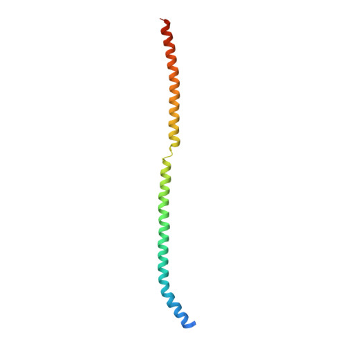
Determining the structural organisation of viral replication complexes and unravelling the impact of infection on cellular homeostasis represent important challenges in virology. This may prove particularly useful when confronted with viruses that pose a significant threat to human health, that appear unique within their family, or for which knowledge is scarce. Among Mononegavirales , bornaviruses (family Bornaviridae ) stand out due to their compact genomes and their nuclear localisation for replication. The recent recognition of the zoonotic potential of several orthobornaviruses has sparked a surge of interest in improving our knowledge on this viral family. In this work, we provide a complete analysis of the structural organisation of Borna disease virus 1 (BoDV-1) phosphoprotein (P), an important cofactor for polymerase activity. Using X-ray diffusion and diffraction experiments, we revealed that BoDV-1 P adopts a long coiled-coil α-helical structure split into two parts by an original β-strand twist motif, which is highly conserved across the members of whole Orthobornavirus genus and may regulate viral replication. In parallel, we used BioID to determine the proximal interactome of P in living cells. We confirmed previously known interactors and identified novel proteins linked to several biological processes such as DNA repair or mRNA metabolism. Altogether, our study provides important structure/function cues, which may improve our understanding of BoDV-1 pathogenesis.
Organizational Affiliation:
Institut de Biologie Structurale (IBS), CEA, CNRS, Université Grenoble Alpes, 38058 Grenoble, France.