8B5S

Crystal Structure of P. aeruginosa WaaG in complex with UDP-glucose


Experimental Data Snapshot

  • Method: X-RAY DIFFRACTION
  • Resolution: 1.95 Å
  • R-Value Free: 0.219 
  • R-Value Work: 0.180 
  • R-Value Observed: 0.182 

wwPDB Validation   3D Report Full Report


Ligand Structure Quality Assessment 


This is version 1.1 of the entry. See complete history


Literature

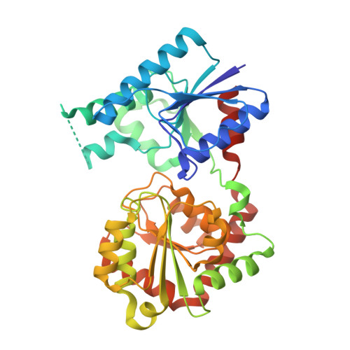
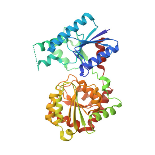
Structural and functional insights into the Pseudomonas aeruginosa glycosyltransferase WaaG and the implications for lipopolysaccharide biosynthesis.

Scaletti, E.R.Pettersson, P.Patrick, J.Shilling, P.J.Westergren, R.G.Daley, D.O.Maler, L.Widmalm, G.Stenmark, P.

(2023) J Biol Chem 299: 105256-105256

  • DOI: https://doi.org/10.1016/j.jbc.2023.105256
  • Primary Citation of Related Structures:  
    8B5Q, 8B5S, 8B62, 8B63

  • PubMed Abstract: 

    The glycosyltransferase WaaG in Pseudomonas aeruginosa (PaWaaG) is involved in the synthesis of the core region of lipopolysaccharides. It is a promising target for developing adjuvants that could help in the uptake of antibiotics. Herein, we have determined structures of PaWaaG in complex with the nucleotide-sugars UDP-glucose, UDP-galactose, and UDP-GalNAc. Structural comparison with the homolog from Escherichia coli (EcWaaG) revealed five key differences in the sugar-binding pocket. Solution-state NMR analysis showed that WT PaWaaG specifically hydrolyzes UDP-GalNAc and unlike EcWaaG, does not hydrolyze UDP-glucose. Furthermore, we found that a PaWaaG mutant (Y97F/T208R/N282A/T283A/T285I) designed to resemble the EcWaaG sugar binding site, only hydrolyzed UDP-glucose, underscoring the importance of the identified amino acids in substrate specificity. However, neither WT PaWaaG nor the PaWaaG mutant capable of hydrolyzing UDP-glucose was able to complement an E. coli ΔwaaG strain, indicating that more remains to be uncovered about the function of PaWaaG in vivo. This structural and biochemical information will guide future structure-based drug design efforts targeting PaWaaG.


  • Organizational Affiliation

    Department of Biochemistry and Biophysics, Stockholm University, Stockholm, Sweden.


Macromolecules
Find similar proteins by:  (by identity cutoff)  |  3D Structure
Entity ID: 1
MoleculeChains Sequence LengthOrganismDetailsImage
UDP-glucose:(Heptosyl) LPS alpha 1,3-glucosyltransferase WaaG374Pseudomonas aeruginosaMutation(s): 0 
Gene Names: waaG
UniProt
Find proteins for Q9HUF6 (Pseudomonas aeruginosa (strain ATCC 15692 / DSM 22644 / CIP 104116 / JCM 14847 / LMG 12228 / 1C / PRS 101 / PAO1))
Explore Q9HUF6 
Go to UniProtKB:  Q9HUF6
Entity Groups  
Sequence Clusters30% Identity50% Identity70% Identity90% Identity95% Identity100% Identity
UniProt GroupQ9HUF6
Sequence Annotations
Expand
  • Reference Sequence
Find similar proteins by:  (by identity cutoff)  |  3D Structure
Entity ID: 2
MoleculeChains Sequence LengthOrganismDetailsImage
UDP-glucose:(Heptosyl) LPS alpha 1,3-glucosyltransferase WaaG370Pseudomonas aeruginosaMutation(s): 0 
Gene Names: waaGPA5010
UniProt
Find proteins for Q9HUF6 (Pseudomonas aeruginosa (strain ATCC 15692 / DSM 22644 / CIP 104116 / JCM 14847 / LMG 12228 / 1C / PRS 101 / PAO1))
Explore Q9HUF6 
Go to UniProtKB:  Q9HUF6
Entity Groups  
Sequence Clusters30% Identity50% Identity70% Identity90% Identity95% Identity100% Identity
UniProt GroupQ9HUF6
Sequence Annotations
Expand
  • Reference Sequence
Small Molecules
Ligands 2 Unique
IDChains Name / Formula / InChI Key2D Diagram3D Interactions
UPG (Subject of Investigation/LOI)
Query on UPG

Download Ideal Coordinates CCD File 
D [auth B]URIDINE-5'-DIPHOSPHATE-GLUCOSE
C15 H24 N2 O17 P2
HSCJRCZFDFQWRP-JZMIEXBBSA-N
UDP
Query on UDP

Download Ideal Coordinates CCD File 
C [auth A]URIDINE-5'-DIPHOSPHATE
C9 H14 N2 O12 P2
XCCTYIAWTASOJW-XVFCMESISA-N
Experimental Data & Validation

Experimental Data

  • Method: X-RAY DIFFRACTION
  • Resolution: 1.95 Å
  • R-Value Free: 0.219 
  • R-Value Work: 0.180 
  • R-Value Observed: 0.182 
  • Space Group: P 1 21 1
Unit Cell:
Length ( Å )Angle ( ˚ )
a = 58.633α = 90
b = 94.426β = 101.41
c = 70.17γ = 90
Software Package:
Software NamePurpose
REFMACrefinement
DIALSdata reduction
Aimlessdata scaling
PHASERphasing

Structure Validation

View Full Validation Report


Ligand Structure Quality Assessment 


Entry History & Funding Information

Deposition Data


Funding OrganizationLocationGrant Number
Swedish Research CouncilSweden--

Revision History  (Full details and data files)

  • Version 1.0: 2023-10-04
    Type: Initial release
  • Version 1.1: 2024-04-17
    Changes: Database references