8AKP

Crystal structure of the catalytic domain of G7048 from Penicillium sumatraense


Experimental Data Snapshot

  • Method: X-RAY DIFFRACTION
  • Resolution: 1.90 Å
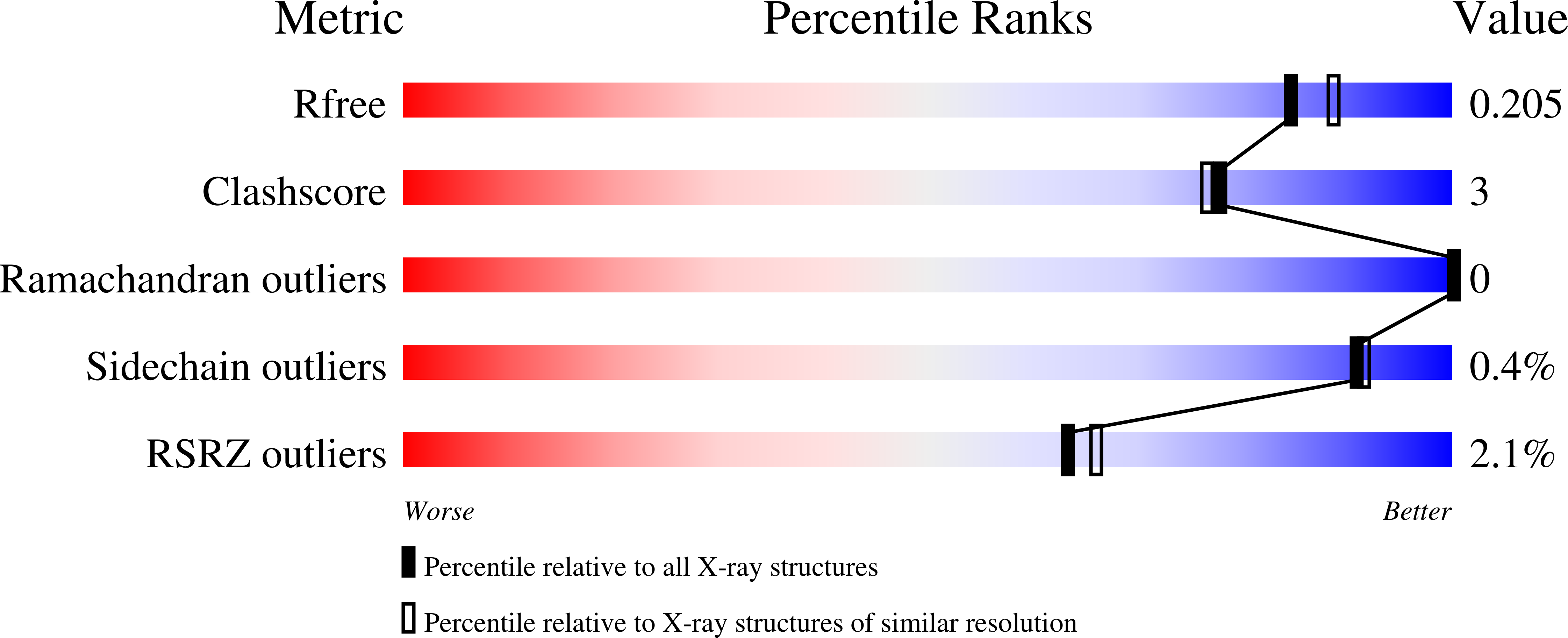
  • R-Value Free: 0.204 
  • R-Value Work: 0.164 
  • R-Value Observed: 0.166 

wwPDB Validation   3D Report Full Report


This is version 1.1 of the entry. See complete history


Literature

Characterization of two 1,3-beta-glucan-modifying enzymes from Penicillium sumatraense reveals new insights into 1,3-beta-glucan metabolism of fungal saprotrophs.

Scafati, V.Troilo, F.Ponziani, S.Giovannoni, M.Scortica, A.Pontiggia, D.Angelucci, F.Di Matteo, A.Mattei, B.Benedetti, M.

(2022) Biotechnol Biofuels Bioprod 15: 138-138

  • DOI: https://doi.org/10.1186/s13068-022-02233-8
  • Primary Citation of Related Structures:  
    8AKP

  • PubMed Abstract: 

    1,3-β-glucan is a polysaccharide widely distributed in the cell wall of several phylogenetically distant organisms, such as bacteria, fungi, plants and microalgae. The presence of highly active 1,3-β-glucanases in fungi evokes the biological question on how these organisms can efficiently metabolize exogenous sources of 1,3-β-glucan without incurring in autolysis.


  • Organizational Affiliation

    Department of Life, Health and Environmental Sciences, University of L'Aquila, 67100, L'Aquila, Italy.


Macromolecules
Find similar proteins by:  (by identity cutoff)  |  3D Structure
Entity ID: 1
MoleculeChains Sequence LengthOrganismDetailsImage
catalytic domain of G7048290Penicillium sumatraenseMutation(s): 0 
Entity Groups  
Sequence Clusters30% Identity50% Identity70% Identity90% Identity95% Identity100% Identity
Sequence Annotations
Expand
  • Reference Sequence
Oligosaccharides

Help

Entity ID: 2
MoleculeChains Length2D Diagram Glycosylation3D Interactions
alpha-D-mannopyranose-(1-3)-[alpha-D-mannopyranose-(1-6)]alpha-D-mannopyranose-(1-6)-[alpha-D-mannopyranose-(1-3)]beta-D-mannopyranose-(1-4)-2-acetamido-2-deoxy-beta-D-glucopyranose-(1-4)-2-acetamido-2-deoxy-beta-D-glucopyranose
B
7N-Glycosylation
Glycosylation Resources
GlyTouCan:  G55220VL
GlyCosmos:  G55220VL
GlyGen:  G55220VL
Small Molecules
Ligands 1 Unique
IDChains Name / Formula / InChI Key2D Diagram3D Interactions
TRS
Query on TRS

Download Ideal Coordinates CCD File 
C [auth A]2-AMINO-2-HYDROXYMETHYL-PROPANE-1,3-DIOL
C4 H12 N O3
LENZDBCJOHFCAS-UHFFFAOYSA-O
Experimental Data & Validation

Experimental Data

  • Method: X-RAY DIFFRACTION
  • Resolution: 1.90 Å
  • R-Value Free: 0.204 
  • R-Value Work: 0.164 
  • R-Value Observed: 0.166 
  • Space Group: P 21 21 2
Unit Cell:
Length ( Å )Angle ( ˚ )
a = 79.22α = 90
b = 87.3β = 90
c = 42.53γ = 90
Software Package:
Software NamePurpose
PHENIXrefinement
XDSdata reduction
Aimlessdata scaling
PHASERphasing

Structure Validation

View Full Validation Report


Entry History & Funding Information

Deposition Data


Funding OrganizationLocationGrant Number
Italian Ministry of EducationItalyPRIN 2017ZBBYNC

Revision History  (Full details and data files)

  • Version 1.0: 2022-12-28
    Type: Initial release
  • Version 1.1: 2024-01-31
    Changes: Data collection, Refinement description