7ZN3

Crystal structure of the light-driven inward proton pump xenorhodopsin BcXeR in the L state at pH 8.2 in the presence of sodium at 100K


Experimental Data Snapshot

  • Method: X-RAY DIFFRACTION
  • Resolution: 1.60 Å
  • R-Value Free: 0.216 
  • R-Value Work: 0.203 
  • R-Value Observed: 0.204 

wwPDB Validation   3D Report Full Report


This is version 1.4 of the entry. See complete history


Literature

Mechanisms of inward transmembrane proton translocation.

Kovalev, K.Tsybrov, F.Alekseev, A.Shevchenko, V.Soloviov, D.Siletsky, S.Bourenkov, G.Agthe, M.Nikolova, M.von Stetten, D.Astashkin, R.Bukhdruker, S.Chizhov, I.Royant, A.Kuzmin, A.Gushchin, I.Rosselli, R.Rodriguez-Valera, F.Ilyinskiy, N.Rogachev, A.Borshchevskiy, V.Schneider, T.R.Bamberg, E.Gordeliy, V.

(2023) Nat Struct Mol Biol 30: 970-979

  • DOI: https://doi.org/10.1038/s41594-023-01020-9
  • Primary Citation of Related Structures:  
    7ZMY, 7ZN0, 7ZN3, 7ZN8, 7ZN9, 7ZNA, 7ZNB, 7ZNC, 7ZND, 7ZNE, 7ZNG, 7ZNH, 7ZNI

  • PubMed Abstract: 

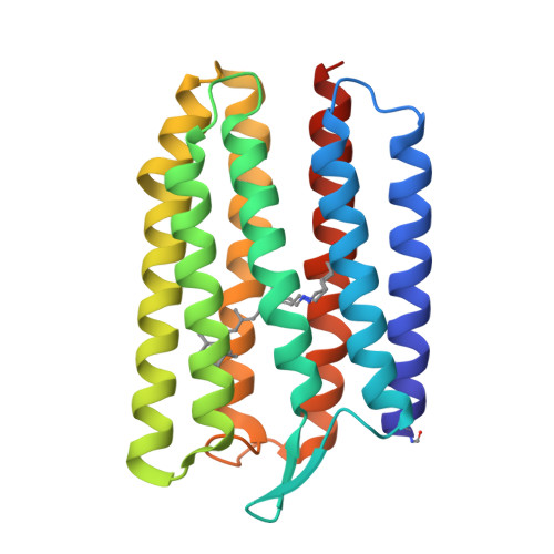
    Proton transport is indispensable for cell life. It is believed that molecular mechanisms of proton movement through different types of proton-conducting molecules have general universal features. However, elucidation of such mechanisms is a challenge. It requires true-atomic-resolution structures of all key proton-conducting states. Here we present a comprehensive function-structure study of a light-driven bacterial inward proton pump, xenorhodopsin, from Bacillus coahuilensis in all major proton-conducting states. The structures reveal that proton translocation is based on proton wires regulated by internal gates. The wires serve as both selectivity filters and translocation pathways for protons. The cumulative results suggest a general concept of proton translocation. We demonstrate the use of serial time-resolved crystallography at a synchrotron source with sub-millisecond resolution for rhodopsin studies, opening the door for principally new applications. The results might also be of interest for optogenetics since xenorhodopsins are the only alternative tools to fire neurons.


  • Organizational Affiliation

    European Molecular Biology Laboratory, Hamburg unit c/o DESY, Hamburg, Germany.


Macromolecules
Find similar proteins by:  (by identity cutoff)  |  3D Structure
Entity ID: 1
MoleculeChains Sequence LengthOrganismDetailsImage
xenorhodopsin
A, B, C
229Bacillus coahuilensisMutation(s): 0 
Entity Groups  
Sequence Clusters30% Identity50% Identity70% Identity90% Identity95% Identity100% Identity
Sequence Annotations
Expand
  • Reference Sequence
Small Molecules
Ligands 4 Unique
IDChains Name / Formula / InChI Key2D Diagram3D Interactions
LFA
Query on LFA

Download Ideal Coordinates CCD File 
AA [auth B]
AB [auth C]
BA [auth B]
BB [auth C]
CB [auth C]
AA [auth B],
AB [auth C],
BA [auth B],
BB [auth C],
CB [auth C],
D [auth A],
DB [auth C],
E [auth A],
EB [auth C],
F [auth A],
FB [auth C],
GB [auth C],
HA [auth B],
HB [auth C],
IA [auth B],
IB [auth C],
JA [auth B],
JB [auth C],
KA [auth B],
KB [auth C],
LA [auth B],
LB [auth C],
M [auth A],
MA [auth B],
N [auth A],
NA [auth B],
O [auth A],
OA [auth B],
P [auth A],
PA [auth B],
Q [auth A],
QA [auth B],
R [auth A],
RA [auth B],
S [auth A],
SA [auth B],
T [auth A],
TA [auth C],
U [auth A],
V [auth A],
W [auth A],
Y [auth B],
Z [auth B],
ZA [auth C]
EICOSANE
C20 H42
CBFCDTFDPHXCNY-UHFFFAOYSA-N
OLA
Query on OLA

Download Ideal Coordinates CCD File 
CA [auth B]
DA [auth B]
EA [auth B]
FA [auth B]
G [auth A]
CA [auth B],
DA [auth B],
EA [auth B],
FA [auth B],
G [auth A],
H [auth A],
I [auth A],
J [auth A],
K [auth A],
UA [auth C],
VA [auth C],
WA [auth C],
XA [auth C]
OLEIC ACID
C18 H34 O2
ZQPPMHVWECSIRJ-KTKRTIGZSA-N
PO4
Query on PO4

Download Ideal Coordinates CCD File 
MB [auth C],
X [auth A]
PHOSPHATE ION
O4 P
NBIIXXVUZAFLBC-UHFFFAOYSA-K
NA (Subject of Investigation/LOI)
Query on NA

Download Ideal Coordinates CCD File 
GA [auth B],
L [auth A],
YA [auth C]
SODIUM ION
Na
FKNQFGJONOIPTF-UHFFFAOYSA-N
Modified Residues  2 Unique
IDChains TypeFormula2D DiagramParent
FME
Query on FME
A, B, C
L-PEPTIDE LINKINGC6 H11 N O3 SMET
LYR
Query on LYR
A, B, C
L-PEPTIDE LINKINGC26 H42 N2 O2LYS
Experimental Data & Validation

Experimental Data

  • Method: X-RAY DIFFRACTION
  • Resolution: 1.60 Å
  • R-Value Free: 0.216 
  • R-Value Work: 0.203 
  • R-Value Observed: 0.204 
  • Space Group: P 21 21 21
Unit Cell:
Length ( Å )Angle ( ˚ )
a = 68.39α = 90
b = 109.49β = 90
c = 119.39γ = 90
Software Package:
Software NamePurpose
PHENIXrefinement
Aimlessdata scaling
PDB_EXTRACTdata extraction
XDSdata reduction
MOLREPphasing

Structure Validation

View Full Validation Report


Entry History & Funding Information

Deposition Data


Funding OrganizationLocationGrant Number
Agence Nationale de la Recherche (ANR)FranceANR-15-CE11-0029-02

Revision History  (Full details and data files)

  • Version 1.0: 2023-05-10
    Type: Initial release
  • Version 1.1: 2023-05-31
    Changes: Derived calculations
  • Version 1.2: 2023-07-12
    Changes: Database references
  • Version 1.3: 2023-07-26
    Changes: Database references
  • Version 1.4: 2024-02-07
    Changes: Data collection, Refinement description