7Z53

Structure of native leukocyte myeloperoxidase in complex with a truncated version (SPIN truncated) of the Staphyloccal Peroxidase Inhibitor SPIN from Staphylococcus aureus


Experimental Data Snapshot

  • Method: X-RAY DIFFRACTION
  • Resolution: 2.28 Å
  • R-Value Free: 0.229 
  • R-Value Work: 0.174 
  • R-Value Observed: 0.177 

wwPDB Validation   3D Report Full Report


Ligand Structure Quality Assessment 


This is version 2.1 of the entry. See complete history


Literature

The staphylococcal inhibitory protein SPIN binds to human myeloperoxidase with picomolar affinity but only dampens halide oxidation.

Leitgeb, U.Furtmuller, P.G.Hofbauer, S.Brito, J.A.Obinger, C.Pfanzagl, V.

(2022) J Biol Chem 298: 102514-102514

  • DOI: https://doi.org/10.1016/j.jbc.2022.102514
  • Primary Citation of Related Structures:  
    7QZR, 7Z53

  • PubMed Abstract: 

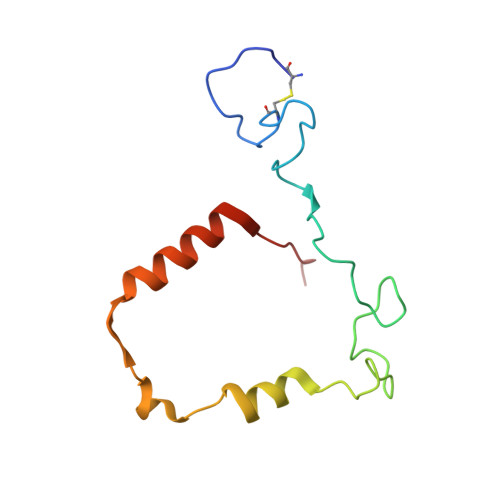
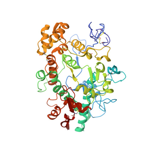
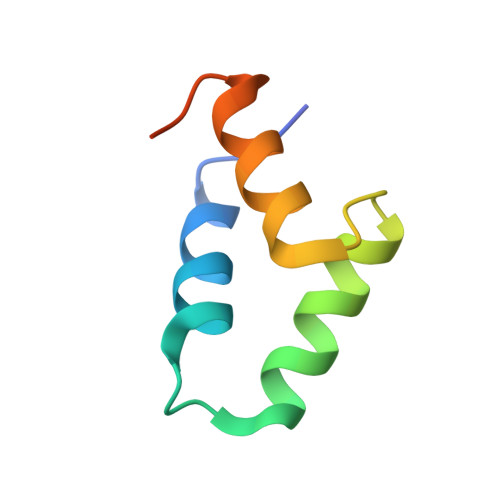
    The heme enzyme myeloperoxidase (MPO) is one of the key players in the neutrophil-mediated killing of invading pathogens as part of the innate immune system. MPO generates antimicrobial oxidants, which indiscriminately and effectively kill phagocytosed pathogens. Staphylococcus aureus, however, is able to escape this fate, in part by secreting a small protein called SPIN (Staphylococcal Peroxidase Inhibitor), which specifically targets and inhibits MPO in a structurally complex manner. Here, we present the first crystal structures of the complex of SPIN-aureus and a truncated version (SPIN-truncated) with mature dimeric leukocyte MPO. We unravel the contributions of the two domains to the kinetics and thermodynamics of SPIN-aureus binding to MPO by using a broad array of complementary biochemical and biophysical methods. The C-terminal "recognition" domain is shown to mediate specific binding to MPO, while interaction of the N-terminal "inhibitory" domain is guided mainly by hydrophobic effects and thus is less sequence dependent. We found that inhibition of MPO is achieved by reducing substrate migration, but SPIN-aureus cannot completely block MPO activity. Its' effectiveness is inversely related to substrate size, with no discernible dependence on other factors. Thus, SPIN-aureus is an extremely high-affinity inhibitor and highly efficient for substrates larger than halogens. As aberrant MPO activity is implicated in a number of chronic inflammatory diseases, SPIN-aureus is the first promising protein inhibitor for specific inhibition of human MPO.


  • Organizational Affiliation

    University of Natural Resources and Life Sciences, Vienna, Department of Chemistry, Institute of Biochemistry, Vienna, Austria.


Macromolecules
Find similar proteins by:  (by identity cutoff)  |  3D Structure
Entity ID: 1
MoleculeChains Sequence LengthOrganismDetailsImage
Myeloperoxidase light chain
A, C, G, I, M
A, C, G, I, M, O, S, U
106Homo sapiensMutation(s): 0 
UniProt & NIH Common Fund Data Resources
Find proteins for P05164 (Homo sapiens)
Explore P05164 
Go to UniProtKB:  P05164
PHAROS:  P05164
GTEx:  ENSG00000005381 
Entity Groups  
Sequence Clusters30% Identity50% Identity70% Identity90% Identity95% Identity100% Identity
UniProt GroupP05164
Sequence Annotations
Expand
  • Reference Sequence
Find similar proteins by:  (by identity cutoff)  |  3D Structure
Entity ID: 2
MoleculeChains Sequence LengthOrganismDetailsImage
Myeloperoxidase heavy chain
B, D, H, J, N
B, D, H, J, N, P, T, V
466Homo sapiensMutation(s): 0 
UniProt & NIH Common Fund Data Resources
Find proteins for P05164 (Homo sapiens)
Explore P05164 
Go to UniProtKB:  P05164
PHAROS:  P05164
GTEx:  ENSG00000005381 
Entity Groups  
Sequence Clusters30% Identity50% Identity70% Identity90% Identity95% Identity100% Identity
UniProt GroupP05164
Sequence Annotations
Expand
  • Reference Sequence
Find similar proteins by:  (by identity cutoff)  |  3D Structure
Entity ID: 3
MoleculeChains Sequence LengthOrganismDetailsImage
Myeloperoxidase inhibitor SPIN
E, F, K, L, Q
E, F, K, L, Q, R, W, X
59Staphylococcus aureusMutation(s): 0 
Gene Names: spnKMZ21_01875
UniProt
Find proteins for Q2G0X2 (Staphylococcus aureus (strain NCTC 8325 / PS 47))
Explore Q2G0X2 
Go to UniProtKB:  Q2G0X2
Entity Groups  
Sequence Clusters30% Identity50% Identity70% Identity90% Identity95% Identity100% Identity
UniProt GroupQ2G0X2
Sequence Annotations
Expand
  • Reference Sequence
Oligosaccharides

Help

Entity ID: 4
MoleculeChains Length2D Diagram Glycosylation3D Interactions
beta-D-mannopyranose-(1-4)-2-acetamido-2-deoxy-beta-D-glucopyranose-(1-4)-2-acetamido-2-deoxy-beta-D-glucopyranoseFA [auth f],
LA [auth l],
OA [auth o],
RA [auth r],
Y
3N-Glycosylation
Glycosylation Resources
GlyTouCan:  G15407YE
GlyCosmos:  G15407YE
GlyGen:  G15407YE
Entity ID: 5
MoleculeChains Length2D Diagram Glycosylation3D Interactions
alpha-D-mannopyranose-(1-6)-beta-D-mannopyranose-(1-4)-2-acetamido-2-deoxy-beta-D-glucopyranose-(1-4)-2-acetamido-2-deoxy-beta-D-glucopyranose
Z
4N-Glycosylation
Glycosylation Resources
GlyTouCan:  G22573RC
GlyCosmos:  G22573RC
GlyGen:  G22573RC
Entity ID: 6
MoleculeChains Length2D Diagram Glycosylation3D Interactions
alpha-D-mannopyranose-(1-3)-[alpha-D-mannopyranose-(1-6)]beta-D-mannopyranose-(1-4)-2-acetamido-2-deoxy-beta-D-glucopyranose-(1-4)-[alpha-L-fucopyranose-(1-6)]2-acetamido-2-deoxy-beta-D-glucopyranose
AA [auth a],
CA [auth c],
EA [auth e],
HA [auth h],
KA [auth k],
AA [auth a],
CA [auth c],
EA [auth e],
HA [auth h],
KA [auth k],
NA [auth n],
QA [auth q],
TA [auth t]
6N-Glycosylation
Glycosylation Resources
GlyTouCan:  G82348BZ
GlyCosmos:  G82348BZ
GlyGen:  G82348BZ
Entity ID: 7
MoleculeChains Length2D Diagram Glycosylation3D Interactions
2-acetamido-2-deoxy-beta-D-glucopyranose-(1-4)-2-acetamido-2-deoxy-beta-D-glucopyranose
BA [auth b],
DA [auth d],
GA [auth g],
MA [auth m],
PA [auth p],
BA [auth b],
DA [auth d],
GA [auth g],
MA [auth m],
PA [auth p],
SA [auth s]
2N-Glycosylation
Glycosylation Resources
GlyTouCan:  G42666HT
GlyCosmos:  G42666HT
GlyGen:  G42666HT
Entity ID: 8
MoleculeChains Length2D Diagram Glycosylation3D Interactions
alpha-D-mannopyranose-(1-3)-beta-D-mannopyranose-(1-4)-2-acetamido-2-deoxy-beta-D-glucopyranose-(1-4)-2-acetamido-2-deoxy-beta-D-glucopyranoseIA [auth i]4N-Glycosylation
Glycosylation Resources
GlyTouCan:  G81315DD
GlyCosmos:  G81315DD
GlyGen:  G81315DD
Entity ID: 9
MoleculeChains Length2D Diagram Glycosylation3D Interactions
alpha-D-mannopyranose-(1-3)-[alpha-D-mannopyranose-(1-6)]beta-D-mannopyranose-(1-4)-2-acetamido-2-deoxy-beta-D-glucopyranose-(1-4)-2-acetamido-2-deoxy-beta-D-glucopyranoseJA [auth j]5N-Glycosylation
Glycosylation Resources
GlyTouCan:  G22768VO
GlyCosmos:  G22768VO
GlyGen:  G22768VO
Small Molecules
Ligands 7 Unique
IDChains Name / Formula / InChI Key2D Diagram3D Interactions
HEC (Subject of Investigation/LOI)
Query on HEC

Download Ideal Coordinates CCD File 
FB [auth G]
JB [auth I]
MB [auth M]
PB [auth O]
UB [auth S]
FB [auth G],
JB [auth I],
MB [auth M],
PB [auth O],
UB [auth S],
VA [auth A],
XB [auth U],
ZA [auth C]
HEME C
C34 H34 Fe N4 O4
HXQIYSLZKNYNMH-LJNAALQVSA-N
NAG
Query on NAG

Download Ideal Coordinates CCD File 
BB [auth D],
CB [auth D],
HB [auth H],
RB [auth P]
2-acetamido-2-deoxy-beta-D-glucopyranose
C8 H15 N O6
OVRNDRQMDRJTHS-FMDGEEDCSA-N
OXL
Query on OXL

Download Ideal Coordinates CCD File 
SB [auth P]OXALATE ION
C2 O4
MUBZPKHOEPUJKR-UHFFFAOYSA-L
EDO
Query on EDO

Download Ideal Coordinates CCD File 
XA [auth B]1,2-ETHANEDIOL
C2 H6 O2
LYCAIKOWRPUZTN-UHFFFAOYSA-N
ACT
Query on ACT

Download Ideal Coordinates CCD File 
DB [auth D]ACETATE ION
C2 H3 O2
QTBSBXVTEAMEQO-UHFFFAOYSA-M
CA
Query on CA

Download Ideal Coordinates CCD File 
AB [auth D]
GB [auth H]
KB [auth J]
NB [auth N]
QB [auth P]
AB [auth D],
GB [auth H],
KB [auth J],
NB [auth N],
QB [auth P],
VB [auth T],
WA [auth B],
YB [auth V]
CALCIUM ION
Ca
BHPQYMZQTOCNFJ-UHFFFAOYSA-N
CL
Query on CL

Download Ideal Coordinates CCD File 
EB [auth G]
IB [auth I]
LB [auth M]
OB [auth O]
TB [auth S]
EB [auth G],
IB [auth I],
LB [auth M],
OB [auth O],
TB [auth S],
UA [auth A],
WB [auth U],
YA [auth C]
CHLORIDE ION
Cl
VEXZGXHMUGYJMC-UHFFFAOYSA-M
Modified Residues  1 Unique
IDChains TypeFormula2D DiagramParent
CSO
Query on CSO
B, D, H, J, N
B, D, H, J, N, P, T, V
L-PEPTIDE LINKINGC3 H7 N O3 SCYS
Experimental Data & Validation

Experimental Data

  • Method: X-RAY DIFFRACTION
  • Resolution: 2.28 Å
  • R-Value Free: 0.229 
  • R-Value Work: 0.174 
  • R-Value Observed: 0.177 
  • Space Group: P 21 21 2
Unit Cell:
Length ( Å )Angle ( ˚ )
a = 257.022α = 90
b = 157.196β = 90
c = 166.56γ = 90
Software Package:
Software NamePurpose
autoPROCdata processing
XDSdata reduction
Aimlessdata scaling
STARANISOdata scaling
MoRDaphasing
Cootmodel building
PHENIXrefinement

Structure Validation

View Full Validation Report


Ligand Structure Quality Assessment 


Entry History & Funding Information

Deposition Data


Funding OrganizationLocationGrant Number
Austrian Science FundAustria33997

Revision History  (Full details and data files)

  • Version 1.0: 2022-10-26
    Type: Initial release
  • Version 1.1: 2022-11-09
    Changes: Database references
  • Version 2.0: 2022-12-07
    Changes: Advisory, Atomic model
  • Version 2.1: 2024-01-31
    Changes: Data collection, Refinement description