7YSE

Crystal structure of E. coli heterotetrameric GlyRS in complex with tRNA


Experimental Data Snapshot

  • Method: X-RAY DIFFRACTION
  • Resolution: 2.91 Å
  • R-Value Free: 0.260 
  • R-Value Work: 0.230 

wwPDB Validation   3D Report Full Report


Ligand Structure Quality Assessment 


This is version 1.2 of the entry. See complete history


Literature

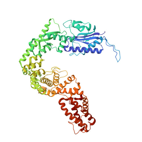
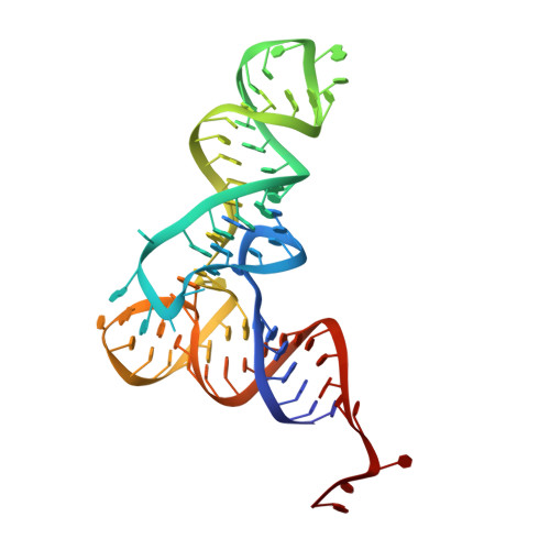
The binding mode of orphan glycyl-tRNA synthetase with tRNA supports the synthetase classification and reveals large domain movements.

Han, L.Luo, Z.Ju, Y.Chen, B.Zou, T.Wang, J.Xu, J.Gu, Q.Yang, X.L.Schimmel, P.Zhou, H.

(2023) Sci Adv 9: eadf1027-eadf1027

  • DOI: https://doi.org/10.1126/sciadv.adf1027
  • Primary Citation of Related Structures:  
    7YSE

  • PubMed Abstract: 

    As a class of essential enzymes in protein translation, aminoacyl-transfer RNA (tRNA) synthetases (aaRSs) are organized into two classes of 10 enzymes each, based on two conserved active site architectures. The (αβ) 2 glycyl-tRNA synthetase (GlyRS) in many bacteria is an orphan aaRS whose sequence and unprecedented X-shaped structure are distinct from those of all other aaRSs, including many other bacterial and all eukaryotic GlyRSs. Here, we report a cocrystal structure to elucidate how the orphan GlyRS kingdom specifically recognizes its substrate tRNA. This structure is sharply different from those of other aaRS-tRNA complexes but conforms to the clash-free, cross-class aaRS-tRNA docking found with conventional structures and reinforces the class-reconstruction paradigm. In addition, noteworthy, the X shape of orphan GlyRS is condensed with the largest known spatial rearrangement needed by aaRSs to capture tRNAs, which suggests potential nonactive site targets for aaRS-directed antibiotics, instead of less differentiated hard-to-drug active site locations.


  • Organizational Affiliation

    Guangdong Provincial Key Laboratory of Chiral Molecule and Drug Discovery, School of Pharmaceutical Sciences, Sun Yat-sen University, Guangzhou 510006, China.


Macromolecules

Find similar proteins by:  (by identity cutoff)  |  3D Structure
Entity ID: 1
MoleculeChains Sequence LengthOrganismDetailsImage
Glycine--tRNA ligase alpha subunit
A, B
303Escherichia coli K-12Mutation(s): 0 
Gene Names: glyQglyS(A)b3560JW3531
EC: 6.1.1.14
UniProt
Find proteins for P00960 (Escherichia coli (strain K12))
Explore P00960 
Go to UniProtKB:  P00960
Entity Groups  
Sequence Clusters30% Identity50% Identity70% Identity90% Identity95% Identity100% Identity
UniProt GroupP00960
Sequence Annotations
Expand
  • Reference Sequence
Find similar proteins by:  (by identity cutoff)  |  3D Structure
Entity ID: 2
MoleculeChains Sequence LengthOrganismDetailsImage
Glycine--tRNA ligase beta subunit
C, D
697Escherichia coli K-12Mutation(s): 0 
Gene Names: glySglyS(B)b3559JW3530
EC: 6.1.1.14
UniProt
Find proteins for P00961 (Escherichia coli (strain K12))
Explore P00961 
Go to UniProtKB:  P00961
Entity Groups  
Sequence Clusters30% Identity50% Identity70% Identity90% Identity95% Identity100% Identity
UniProt GroupP00961
Sequence Annotations
Expand
  • Reference Sequence
Find similar nucleic acids by:  (by identity cutoff)  |  3D Structure
Entity ID: 3
MoleculeChains LengthOrganismImage
RNA (76-MER)
E, F
76Escherichia coli
Sequence Annotations
Expand
  • Reference Sequence
Small Molecules
Ligands 2 Unique
IDChains Name / Formula / InChI Key2D Diagram3D Interactions
JPO (Subject of Investigation/LOI)
Query on JPO

Download Ideal Coordinates CCD File 
G [auth A],
I [auth B]
[(2R,3S,4R,5R)-5-(6-azanyl-2-chloranyl-purin-9-yl)-3,4-bis(oxidanyl)oxolan-2-yl]methyl N-(2-azanylethanoyl)sulfamate
C12 H16 Cl N7 O7 S
GSOIQTQOUNQJFI-TZQXKBMNSA-N
MG
Query on MG

Download Ideal Coordinates CCD File 
H [auth A]
J [auth B]
K [auth C]
L [auth C]
M [auth D]
H [auth A],
J [auth B],
K [auth C],
L [auth C],
M [auth D],
N [auth E]
MAGNESIUM ION
Mg
JLVVSXFLKOJNIY-UHFFFAOYSA-N
Experimental Data & Validation

Experimental Data

  • Method: X-RAY DIFFRACTION
  • Resolution: 2.91 Å
  • R-Value Free: 0.260 
  • R-Value Work: 0.230 
  • Space Group: P 2 2 21
Unit Cell:
Length ( Å )Angle ( ˚ )
a = 72.71α = 90
b = 162.123β = 90
c = 324.09γ = 90
Software Package:
Software NamePurpose
REFMACrefinement
autoPROCdata reduction
Aimlessdata scaling
MOLREPphasing

Structure Validation

View Full Validation Report


Ligand Structure Quality Assessment 


Entry History & Funding Information

Deposition Data


Funding OrganizationLocationGrant Number
National Natural Science Foundation of China (NSFC)China22177140

Revision History  (Full details and data files)

  • Version 1.0: 2023-02-01
    Type: Initial release
  • Version 1.1: 2023-02-22
    Changes: Database references, Structure summary
  • Version 1.2: 2023-11-29
    Changes: Data collection, Refinement description