Structure-based rational design of a short-chain dehydrogenase/reductase for improving activity toward mycotoxin patulin.
Dai, L., Li, H., Huang, J.W., Hu, Y., He, M., Yang, Y., Min, J., Guo, R.T., Chen, C.C.(2022) Int J Biol Macromol 222: 421-428
- PubMed: 36176222
- DOI: https://doi.org/10.1016/j.ijbiomac.2022.09.121
- Primary Citation of Related Structures:
7XWH, 7XWI, 7XWJ, 7XWK, 7XWL, 7XWM, 7XWN - PubMed Abstract:
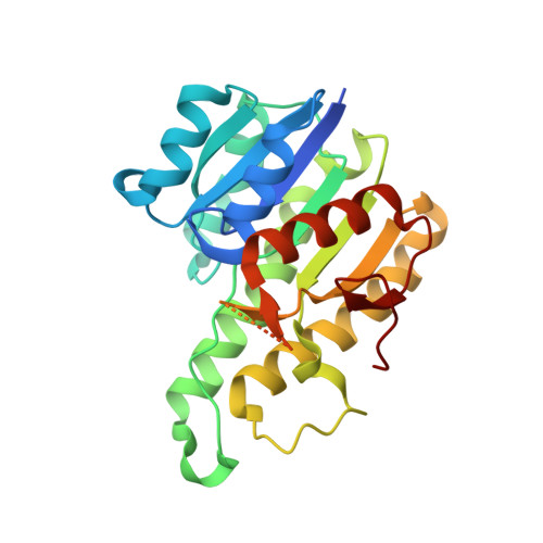
Patulin is a fatal mycotoxin that is widely detected in drinking water and fruit-derived products contaminated by diverse filamentous fungi. CgSDR from Candida guilliermondii represents the first NADPH-dependent short-chain dehydrogenase/reductase that catalyzes the reduction of patulin to the nontoxic E-ascladiol. To elucidate the catalytic mechanism of CgSDR, we solved its crystal structure in complex with cofactor and substrate. Structural analyses indicate that patulin is situated in a hydrophobic pocket adjacent to the cofactor, with the hemiacetal ring orienting toward the nicotinamide moiety of NADPH. In addition, we conducted structure-guided engineering to modify substrate-binding residue V187 and obtained variant V187F, V187K and V187W, whose catalytic activity was elevated by 3.9-, 2.2- and 1.7-fold, respectively. The crystal structures of CgSDR variants suggest that introducing additional aromatic stacking or hydrogen-bonding interactions to bind the lactone ring of patulin might account for the observed enhanced activity. These results illustrate the catalytic mechanism of SDR-mediated patulin detoxification for the first time and provide the upgraded variants that exhibit tremendous potentials in industrial applications.
Organizational Affiliation:
State Key Laboratory of Biocatalysis and Enzyme Engineering, Hubei Hongshan Laboratory, Hubei Collaborative Innovation Center for Green Transformation of Bio-Resources, Hubei Key Laboratory of Industrial Biotechnology, School of Life Sciences, Hubei University, Wuhan, 430062, PR China.