7UWI

Structure of beta-catenin in complex with FP01567, a Helicon Polypeptide


Experimental Data Snapshot

  • Method: X-RAY DIFFRACTION
  • Resolution: 2.32 Å
  • R-Value Free: 0.278 
  • R-Value Work: 0.236 
  • R-Value Observed: 0.238 

wwPDB Validation   3D Report Full Report


Ligand Structure Quality Assessment 


This is version 1.1 of the entry. See complete history


Literature

De novo mapping of alpha-helix recognition sites on protein surfaces using unbiased libraries.

Li, K.Tokareva, O.S.Thomson, T.M.Wahl, S.C.T.Travaline, T.L.Ramirez, J.D.Choudary, S.K.Agarwal, S.Walkup 4th, W.G.Olsen, T.J.Brennan, M.J.Verdine, G.L.McGee, J.H.

(2022) Proc Natl Acad Sci U S A 119: e2210435119-e2210435119

  • DOI: https://doi.org/10.1073/pnas.2210435119
  • Primary Citation of Related Structures:  
    7UWI, 7UWO, 7UX5, 7UXI, 7UXJ, 7UXK, 7UXM, 7UXN, 7UXO, 7UXP, 7UXQ, 7UY2, 7UYJ, 7UYK

  • PubMed Abstract: 

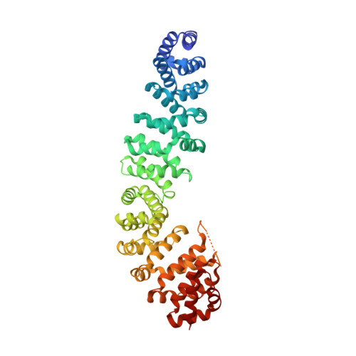
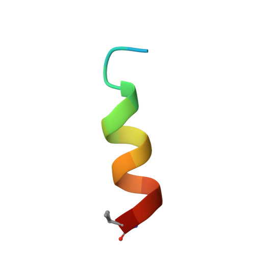
    The α-helix is one of the most common protein surface recognition motifs found in nature, and its unique amide-cloaking properties also enable α-helical polypeptide motifs to exist in membranes. Together, these properties have inspired the development of α-helically constrained (Helicon) therapeutics that can enter cells and bind targets that have been considered "undruggable", such as protein-protein interactions. To date, no general method for discovering α-helical binders to proteins has been reported, limiting Helicon drug discovery to only those proteins with previously characterized α-helix recognition sites, and restricting the starting chemical matter to those known α-helical binders. Here, we report a general and rapid screening method to empirically map the α-helix binding sites on a broad range of target proteins in parallel using large, unbiased Helicon phage display libraries and next-generation sequencing. We apply this method to screen six structurally diverse protein domains, only one of which had been previously reported to bind isolated α-helical peptides, discovering 20 families that collectively comprise several hundred individual Helicons. Analysis of 14 X-ray cocrystal structures reveals at least nine distinct α-helix recognition sites across these six proteins, and biochemical and biophysical studies show that these Helicons can block protein-protein interactions, inhibit enzymatic activity, induce conformational rearrangements, and cause protein dimerization. We anticipate that this method will prove broadly useful for the study of protein recognition and for the development of both biochemical tools and therapeutics for traditionally challenging protein targets.


  • Organizational Affiliation

    FOG Pharmaceuticals Inc., Cambridge, MA 02140.


Macromolecules
Find similar proteins by:  (by identity cutoff)  |  3D Structure
Entity ID: 1
MoleculeChains Sequence LengthOrganismDetailsImage
Catenin beta-1521Homo sapiensMutation(s): 0 
Gene Names: CTNNB1CTNNBOK/SW-cl.35PRO2286
UniProt & NIH Common Fund Data Resources
Find proteins for P35222 (Homo sapiens)
Explore P35222 
Go to UniProtKB:  P35222
PHAROS:  P35222
GTEx:  ENSG00000168036 
Entity Groups  
Sequence Clusters30% Identity50% Identity70% Identity90% Identity95% Identity100% Identity
UniProt GroupP35222
Sequence Annotations
Expand
  • Reference Sequence

Find similar proteins by:  Sequence   |   3D Structure  

Entity ID: 2
MoleculeChains Sequence LengthOrganismDetailsImage
Helicon Polypeptide FP01567B [auth F]16synthetic constructMutation(s): 0 
Entity Groups  
Sequence Clusters30% Identity50% Identity70% Identity90% Identity95% Identity100% Identity
Sequence Annotations
Expand
  • Reference Sequence
Small Molecules
Ligands 3 Unique
IDChains Name / Formula / InChI Key2D Diagram3D Interactions
WHL (Subject of Investigation/LOI)
Query on WHL

Download Ideal Coordinates CCD File 
J [auth F]N,N'-(1,4-phenylene)diacetamide
C10 H12 N2 O2
KVEDKKLZCJBVNP-UHFFFAOYSA-N
PO4
Query on PO4

Download Ideal Coordinates CCD File 
C [auth A]PHOSPHATE ION
O4 P
NBIIXXVUZAFLBC-UHFFFAOYSA-K
GOL
Query on GOL

Download Ideal Coordinates CCD File 
D [auth A]
E [auth A]
F [auth A]
G [auth A]
H [auth A]
D [auth A],
E [auth A],
F [auth A],
G [auth A],
H [auth A],
I [auth A]
GLYCEROL
C3 H8 O3
PEDCQBHIVMGVHV-UHFFFAOYSA-N
Experimental Data & Validation

Experimental Data

  • Method: X-RAY DIFFRACTION
  • Resolution: 2.32 Å
  • R-Value Free: 0.278 
  • R-Value Work: 0.236 
  • R-Value Observed: 0.238 
  • Space Group: P 21 21 21
Unit Cell:
Length ( Å )Angle ( ˚ )
a = 49.68α = 90
b = 91.81β = 90
c = 113.35γ = 90
Software Package:
Software NamePurpose
PHENIXrefinement
PDB_EXTRACTdata extraction
XDSdata scaling
XDSdata reduction
PHASERphasing

Structure Validation

View Full Validation Report


Ligand Structure Quality Assessment 


Entry History & Funding Information

Deposition Data


Funding OrganizationLocationGrant Number
Other privateUnited States--

Revision History  (Full details and data files)

  • Version 1.0: 2022-12-28
    Type: Initial release
  • Version 1.1: 2023-10-25
    Changes: Data collection, Refinement description