Characterization of the Serpentine Adeno-Associated Virus (SAAV) Capsid Structure: Receptor Interactions and Antigenicity.
Mietzsch, M., Hull, J.A., Makal, V.E., Jimenez Ybargollin, A., Yu, J.C., McKissock, K., Bennett, A., Penzes, J., Lins-Austin, B., Yu, Q., Chipman, P., Bhattacharya, N., Sousa, D., Strugatsky, D., Tijssen, P., McKenna, R., Agbandje-McKenna, M.(2022) J Virol 96: e0033522-e0033522
- PubMed: 35532224
- DOI: https://doi.org/10.1128/jvi.00335-22
- Primary Citation of Related Structures:
7U94, 7U95, 7U96, 7U97 - PubMed Abstract:
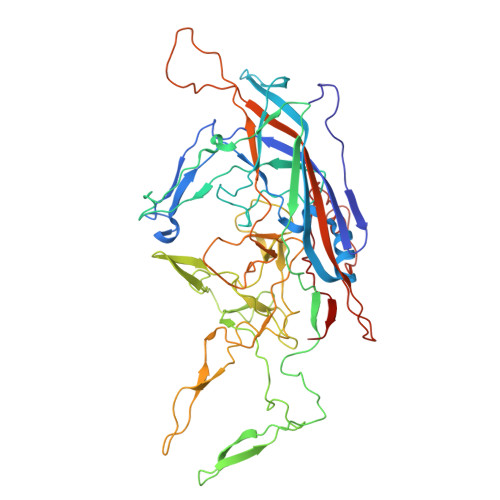
Adeno-associated viruses (AAVs) are being developed as clinical gene therapy vectors. One issue undermining their broad use in the clinical setting is the high prevalence of circulating antibodies in the general population capable of neutralizing AAV vectors. Hence, there is a need for AAV vectors that can evade the preexisting immune response. One possible source of human naive vectors are AAVs that do not disseminate in the primate population, and one such example is serpentine AAV (SAAV). This study characterizes the structural and biophysical properties of the SAAV capsid and its receptor interactions and antigenicity. Single particle cryo-electron microscopy (cryo-EM) and thermal stability studies were conducted to characterize the SAAV capsid structure at pH 7.4, 6.0, 5.5, and 4.0, conditions experienced during cellular trafficking. Cell binding assays using Chinese hamster ovary (CHO) cell lines identified terminal sialic acid as the primary attachment receptor for SAAV similar to AAV1, 4, 5, and 6. The binding site of sialic acid to the SAAV capsid was mapped near the 2-fold axis toward the 2/5-fold wall, in a different location than AAV1, 4, 5, and 6. Towards determining the SAAV capsid antigenicity native immunodot blots showed that SAAV evades AAV serotype-specific mouse monoclonal antibodies. However, despite its reptilian origin, it was recognized by ~25% of 50 human sera tested, likely due to the presence of cross-reactive antibodies. These findings will inform future gene delivery applications using SAAV-based vectors and further aid the structural characterization and annotation of the repertoire of available AAV capsids. IMPORTANCE AAVs are widely studied therapeutic gene delivery vectors. However, preexisting antibodies and their detrimental effect on therapeutic efficacy are a primary challenge encountered during clinical trials. In order to circumvent preexisting neutralizing antibodies targeting mammalian AAV capsids, serpentine AAV (SAAV) was evaluated as a potential alternative to existing mammalian therapeutic vectors. The SAAV capsid was found to be thermostable at a wide range of environmental pH conditions, and its structure showed conservation of the core capsid topology but displays high structural variability on the surface. At the same time, it binds to a common receptor, sialic acid, that is also utilized by other AAVs already being utilized in gene therapy trials. Contrary to the initial hypothesis, SAAV capsids were recognized by one in four human sera tested, pointing to conserved amino acids around the 5-fold region as epitopes for cross-reacting antibodies.
Organizational Affiliation:
University of Floridagrid.15276.37, Department of Biochemistry & Molecular Biology, Gainesville, Florida, USA.