7U02

Structure of the C. crescentus DriD C-domain bound to ssDNA


Experimental Data Snapshot

  • Method: X-RAY DIFFRACTION
  • Resolution: 2.48 Å
  • R-Value Free: 0.279 
  • R-Value Work: 0.230 
  • R-Value Observed: 0.234 

wwPDB Validation   3D Report Full Report


Ligand Structure Quality Assessment 


This is version 1.3 of the entry. See complete history


Literature

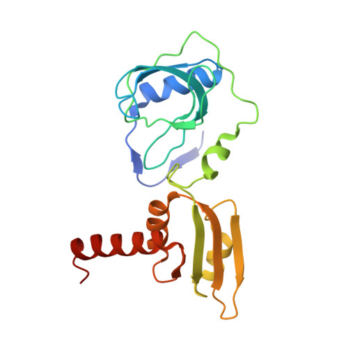
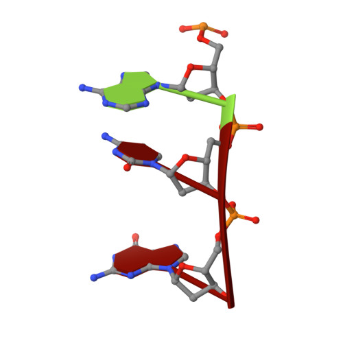
ssDNA is an allosteric regulator of the C. crescentus SOS-independent DNA damage response transcription activator, DriD.

Gozzi, K.Salinas, R.Nguyen, V.D.Laub, M.T.Schumacher, M.A.

(2022) Genes Dev 36: 618-633

  • DOI: https://doi.org/10.1101/gad.349541.122
  • Primary Citation of Related Structures:  
    7TZV, 7U02

  • PubMed Abstract: 

    DNA damage repair systems are critical for genomic integrity. However, they must be coordinated with DNA replication and cell division to ensure accurate genomic transmission. In most bacteria, this coordination is mediated by the SOS response through LexA, which triggers a halt in cell division until repair is completed. Recently, an SOS-independent damage response system was revealed in Caulobacter crescentus. This pathway is controlled by the transcription activator, DriD, but how DriD senses and signals DNA damage is unknown. To address this question, we performed biochemical, cellular, and structural studies. We show that DriD binds a specific promoter DNA site via its N-terminal HTH domain to activate transcription of genes, including the cell division inhibitor didA A structure of the C-terminal portion of DriD revealed a WYL motif domain linked to a WCX dimerization domain. Strikingly, we found that DriD binds ssDNA between the WYL and WCX domains. Comparison of apo and ssDNA-bound DriD structures reveals that ssDNA binding orders and orients the DriD domains, indicating a mechanism for ssDNA-mediated operator DNA binding activation. Biochemical and in vivo studies support the structural model. Our data thus reveal the molecular mechanism underpinning an SOS-independent DNA damage repair pathway.


  • Organizational Affiliation

    Department of Biology, Massachusetts Institute of Technology. Cambridge, Massachusetts 02139, USA.


Macromolecules

Find similar proteins by:  (by identity cutoff)  |  3D Structure
Entity ID: 1
MoleculeChains Sequence LengthOrganismDetailsImage
WYL domain-containing proteinA [auth B],
C [auth M]
195Caulobacter vibrioides CB15Mutation(s): 0 
Gene Names: CC_1095
UniProt
Find proteins for Q9A999 (Caulobacter vibrioides (strain ATCC 19089 / CB15))
Explore Q9A999 
Go to UniProtKB:  Q9A999
Entity Groups  
Sequence Clusters30% Identity50% Identity70% Identity90% Identity95% Identity100% Identity
UniProt GroupQ9A999
Sequence Annotations
Expand
  • Reference Sequence

Find similar nucleic acids by:  Sequence   |   3D Structure  

Entity ID: 2
MoleculeChains LengthOrganismImage
DNA (5'-D(P*AP*CP*G)-3')B [auth H]3synthetic construct
Sequence Annotations
Expand
  • Reference Sequence
Experimental Data & Validation

Experimental Data

  • Method: X-RAY DIFFRACTION
  • Resolution: 2.48 Å
  • R-Value Free: 0.279 
  • R-Value Work: 0.230 
  • R-Value Observed: 0.234 
  • Space Group: P 31 2 1
Unit Cell:
Length ( Å )Angle ( ˚ )
a = 87.4α = 90
b = 87.4β = 90
c = 139.6γ = 120
Software Package:
Software NamePurpose
PHENIXrefinement
XDSdata reduction
SCALAdata scaling
PHASERphasing
PDB_EXTRACTdata extraction

Structure Validation

View Full Validation Report


Ligand Structure Quality Assessment 


Entry History & Funding Information

Deposition Data


Funding OrganizationLocationGrant Number
National Institutes of Health/National Institute of General Medical Sciences (NIH/NIGMS)United StatesR35GM130290

Revision History  (Full details and data files)

  • Version 1.0: 2022-06-01
    Type: Initial release
  • Version 1.1: 2022-06-08
    Changes: Database references
  • Version 1.2: 2022-06-22
    Changes: Database references
  • Version 1.3: 2024-02-21
    Changes: Data collection