7T6F

Structure of active Janus Kinase (JAK) dimer complexed with cytokine receptor intracellular domain


Experimental Data Snapshot

  • Method: ELECTRON MICROSCOPY
  • Resolution: 3.60 Å
  • Aggregation State: PARTICLE 
  • Reconstruction Method: SINGLE PARTICLE 

wwPDB Validation   3D Report Full Report


This is version 1.3 of the entry. See complete history


Literature

Structure of a Janus kinase cytokine receptor complex reveals the basis for dimeric activation.

Glassman, C.R.Tsutsumi, N.Saxton, R.A.Lupardus, P.J.Jude, K.M.Garcia, K.C.

(2022) Science 376: 163-169

  • DOI: https://doi.org/10.1126/science.abn8933
  • Primary Citation of Related Structures:  
    7T6F

  • PubMed Abstract: 

    Cytokines signal through cell surface receptor dimers to initiate activation of intracellular Janus kinases (JAKs). We report the 3.6-angstrom-resolution cryo-electron microscopy structure of full-length JAK1 complexed with a cytokine receptor intracellular domain Box1 and Box2 regions captured as an activated homodimer bearing the valine→phenylalanine (VF) mutation prevalent in myeloproliferative neoplasms. The seven domains of JAK1 form an extended structural unit, the dimerization of which is mediated by close-packing of the pseudokinase (PK) domains from the monomeric subunits. The oncogenic VF mutation lies within the core of the JAK1 PK interdimer interface, enhancing packing complementarity to facilitate ligand-independent activation. The carboxy-terminal tyrosine kinase domains are poised for transactivation and to phosphorylate the receptor STAT (signal transducer and activator of transcription)-recruiting motifs projecting from the overhanging FERM (four-point-one, ezrin, radixin, moesin)-SH2 (Src homology 2)-domains. Mapping of constitutively active JAK mutants supports a two-step allosteric activation mechanism and reveals opportunities for selective therapeutic targeting of oncogenic JAK signaling.


  • Organizational Affiliation

    Department of Molecular and Cellular Physiology, Stanford University School of Medicine, Stanford, CA 94305, USA.


Macromolecules
Find similar proteins by:  (by identity cutoff)  |  3D Structure
Entity ID: 1
MoleculeChains Sequence LengthOrganismDetailsImage
Tyrosine-protein kinaseA,
D [auth B]
1,173Mus musculusMutation(s): 1 
Gene Names: Jak1
EC: 2.7.10.2
UniProt
Find proteins for P52332 (Mus musculus)
Explore P52332 
Go to UniProtKB:  P52332
Entity Groups  
Sequence Clusters30% Identity50% Identity70% Identity90% Identity95% Identity100% Identity
UniProt GroupP52332
Sequence Annotations
Expand
  • Reference Sequence
Find similar proteins by:  (by identity cutoff)  |  3D Structure
Entity ID: 2
MoleculeChains Sequence LengthOrganismDetailsImage
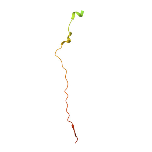
Interferon lambda receptor 1B [auth C],
C [auth D]
85Mus musculusMutation(s): 0 
Gene Names: Ifnlr1Il28ra
UniProt & NIH Common Fund Data Resources
Find proteins for Q8CGK5 (Mus musculus)
Explore Q8CGK5 
Go to UniProtKB:  Q8CGK5
IMPC:  MGI:2429859
Entity Groups  
Sequence Clusters30% Identity50% Identity70% Identity90% Identity95% Identity100% Identity
UniProt GroupQ8CGK5
Sequence Annotations
Expand
  • Reference Sequence
Experimental Data & Validation

Experimental Data

  • Method: ELECTRON MICROSCOPY
  • Resolution: 3.60 Å
  • Aggregation State: PARTICLE 
  • Reconstruction Method: SINGLE PARTICLE 
EM Software:
TaskSoftware PackageVersion
MODEL REFINEMENTPHENIX
RECONSTRUCTIONcryoSPARC3.3

Structure Validation

View Full Validation Report


Entry History & Funding Information

Deposition Data


Funding OrganizationLocationGrant Number
National Institutes of Health/National Institute Of Allergy and Infectious Diseases (NIH/NIAID)United StatesR37AI051321
Howard Hughes Medical Institute (HHMI)United States--
Ludwig Institute for Cancer Research (LICR)United States--

Revision History  (Full details and data files)

  • Version 1.0: 2022-03-16
    Type: Initial release
  • Version 1.1: 2022-03-23
    Changes: Database references
  • Version 1.2: 2022-04-20
    Changes: Database references
  • Version 1.3: 2024-02-28
    Changes: Data collection