7T1A

Rev1 Ternary Complex with dATP and Ca2+


Experimental Data Snapshot

  • Method: X-RAY DIFFRACTION
  • Resolution: 1.81 Å
  • R-Value Free: 0.230 
  • R-Value Work: 0.193 
  • R-Value Observed: 0.195 

wwPDB Validation   3D Report Full Report


Ligand Structure Quality Assessment 


This is version 1.2 of the entry. See complete history


Literature

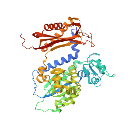
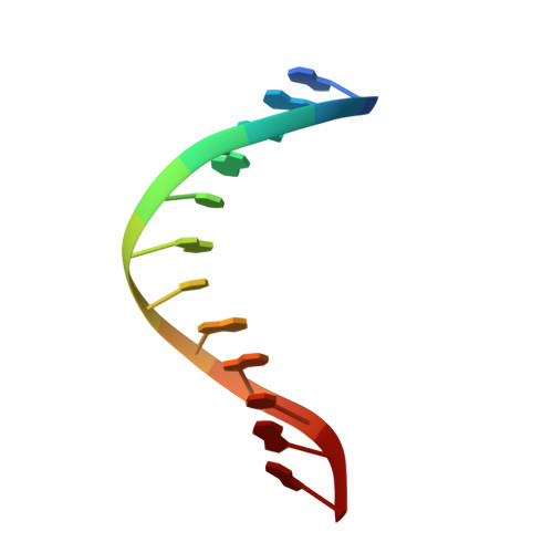
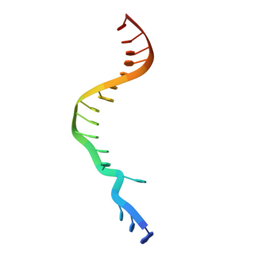
Mechanism of nucleotide discrimination by the translesion synthesis polymerase Rev1.

Weaver, T.M.Click, T.H.Khoang, T.H.Todd Washington, M.Agarwal, P.K.Freudenthal, B.D.

(2022) Nat Commun 13: 2876-2876

  • DOI: https://doi.org/10.1038/s41467-022-30577-0
  • Primary Citation of Related Structures:  
    7T18, 7T19, 7T1A, 7T1B

  • PubMed Abstract: 

    Rev1 is a translesion DNA synthesis (TLS) polymerase involved in the bypass of adducted-guanine bases and abasic sites during DNA replication. During damage bypass, Rev1 utilizes a protein-template mechanism of DNA synthesis, where the templating DNA base is evicted from the Rev1 active site and replaced by an arginine side chain that preferentially binds incoming dCTP. Here, we utilize X-ray crystallography and molecular dynamics simulations to obtain structural insight into the dCTP specificity of Rev1. We show the Rev1 R324 protein-template forms sub-optimal hydrogen bonds with incoming dTTP, dGTP, and dATP that prevents Rev1 from adopting a catalytically competent conformation. Additionally, we show the Rev1 R324 protein-template forms optimal hydrogen bonds with incoming rCTP. However, the incoming rCTP adopts an altered sugar pucker, which prevents the formation of a catalytically competent Rev1 active site. This work provides novel insight into the mechanisms for nucleotide discrimination by the TLS polymerase Rev1.


  • Organizational Affiliation

    Department of Biochemistry and Molecular Biology, University of Kansas Medical Center, Kansas City, Kansas, 66160, USA.


Macromolecules

Find similar proteins by:  (by identity cutoff)  |  3D Structure
Entity ID: 3
MoleculeChains Sequence LengthOrganismDetailsImage
DNA repair protein REV1C [auth A]451Saccharomyces cerevisiaeMutation(s): 0 
Gene Names: REV1YOR346WO6339
EC: 2.7.7
UniProt
Find proteins for P12689 (Saccharomyces cerevisiae (strain ATCC 204508 / S288c))
Explore P12689 
Go to UniProtKB:  P12689
Entity Groups  
Sequence Clusters30% Identity50% Identity70% Identity90% Identity95% Identity100% Identity
UniProt GroupP12689
Sequence Annotations
Expand
  • Reference Sequence

Find similar nucleic acids by:  Sequence   |   3D Structure  

Entity ID: 1
MoleculeChains LengthOrganismImage
DNA (5'-D(*GP*GP*GP*GP*TP*GP*TP*GP*GP*TP*AP*G)-3')A [auth P]12synthetic construct
Sequence Annotations
Expand
  • Reference Sequence

Find similar nucleic acids by:  Sequence   |   3D Structure  

Entity ID: 2
MoleculeChains LengthOrganismImage
DNA (5'-D(*AP*TP*CP*GP*CP*TP*AP*CP*CP*AP*CP*AP*CP*CP*CP*C)-3')B [auth T]17synthetic construct
Sequence Annotations
Expand
  • Reference Sequence
Experimental Data & Validation

Experimental Data

  • Method: X-RAY DIFFRACTION
  • Resolution: 1.81 Å
  • R-Value Free: 0.230 
  • R-Value Work: 0.193 
  • R-Value Observed: 0.195 
  • Space Group: P 21 21 21
Unit Cell:
Length ( Å )Angle ( ˚ )
a = 62.136α = 90
b = 73.336β = 90
c = 115.446γ = 90
Software Package:
Software NamePurpose
PHENIXrefinement
HKL-2000data reduction
HKL-2000data scaling
PHENIXphasing

Structure Validation

View Full Validation Report


Ligand Structure Quality Assessment 


Entry History & Funding Information

Deposition Data


Funding OrganizationLocationGrant Number
National Institutes of Health/National Institute of General Medical Sciences (NIH/NIGMS)United States--

Revision History  (Full details and data files)

  • Version 1.0: 2022-05-25
    Type: Initial release
  • Version 1.1: 2022-06-08
    Changes: Database references
  • Version 1.2: 2023-10-18
    Changes: Data collection, Refinement description