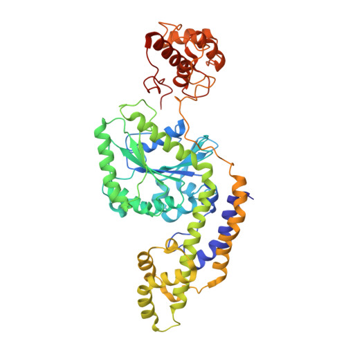
Cryo-electron tomography reveals structural insights into the membrane remodeling mode of dynamin-like EHD filaments.
Melo, A.A., Sprink, T., Noel, J.K., Vazquez-Sarandeses, E., van Hoorn, C., Mohd, S., Loerke, J., Spahn, C.M.T., Daumke, O.(2022) Nat Commun 13: 7641-7641
- PubMed: 36496453
- DOI: https://doi.org/10.1038/s41467-022-35164-x
- Primary Citation of Related Structures:
7SOX - PubMed Abstract:
Eps15-homology domain containing proteins (EHDs) are eukaryotic, dynamin-related ATPases involved in cellular membrane trafficking. They oligomerize on membranes into filaments that induce membrane tubulation. While EHD crystal structures in open and closed conformations were previously reported, little structural information is available for the membrane-bound oligomeric form. Consequently, mechanistic insights into the membrane remodeling mechanism have remained sparse. Here, by using cryo-electron tomography and subtomogram averaging, we determined structures of nucleotide-bound EHD4 filaments on membrane tubes of various diameters at an average resolution of 7.6 Å. Assembly of EHD4 is mediated via interfaces in the G-domain and the helical domain. The oligomerized EHD4 structure resembles the closed conformation, where the tips of the helical domains protrude into the membrane. The variation in filament geometry and tube radius suggests a spontaneous filament curvature of approximately 1/70 nm -1 . Combining the available structural and functional data, we suggest a model for EHD-mediated membrane remodeling.
Organizational Affiliation:
Max-Delbrück-Center for Molecular Medicine in the Helmholtz Association, Structural Biology, Robert-Rössle-Straße 10, Berlin, Germany. arthur.melo@ucsf.edu.