7S8W

Amycolatopsis sp. T-1-60 N-succinylamino acid racemase/o-succinylbenzoate synthase R266Q mutant in complex with N-succinylphenylglycine


Experimental Data Snapshot

  • Method: X-RAY DIFFRACTION
  • Resolution: 2.90 Å
  • R-Value Free: 0.215 
  • R-Value Work: 0.170 
  • R-Value Observed: 0.172 

wwPDB Validation   3D Report Full Report


Ligand Structure Quality Assessment 


This is version 1.3 of the entry. See complete history


Literature

Second-Shell Amino Acid R266 Helps Determine N -Succinylamino Acid Racemase Reaction Specificity in Promiscuous N -Succinylamino Acid Racemase/ o -Succinylbenzoate Synthase Enzymes.

Truong, D.P.Rousseau, S.Machala, B.W.Huddleston, J.P.Zhu, M.Hull, K.G.Romo, D.Raushel, F.M.Sacchettini, J.C.Glasner, M.E.

(2021) Biochemistry 60: 3829-3840

  • DOI: https://doi.org/10.1021/acs.biochem.1c00627
  • Primary Citation of Related Structures:  
    7S8W

  • PubMed Abstract: 

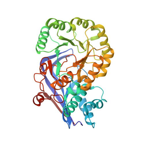
    Catalytic promiscuity is the coincidental ability to catalyze nonbiological reactions in the same active site as the native biological reaction. Several lines of evidence show that catalytic promiscuity plays a role in the evolution of new enzyme functions. Thus, studying catalytic promiscuity can help identify structural features that predispose an enzyme to evolve new functions. This study identifies a potentially preadaptive residue in a promiscuous N -succinylamino acid racemase/ o -succinylbenzoate synthase (NSAR/OSBS) enzyme from Amycolatopsis sp. T-1-60. This enzyme belongs to a branch of the OSBS family which includes many catalytically promiscuous NSAR/OSBS enzymes. R266 is conserved in all members of the NSAR/OSBS subfamily. However, the homologous position is usually hydrophobic in other OSBS subfamilies, whose enzymes lack NSAR activity. The second-shell amino acid R266 is close to the catalytic acid/base K263, but it does not contact the substrate, suggesting that R266 could affect the catalytic mechanism. Mutating R266 to glutamine in Amycolatopsis NSAR/OSBS profoundly reduces NSAR activity but moderately reduces OSBS activity. This is due to a 1000-fold decrease in the rate of proton exchange between the substrate and the general acid/base catalyst K263. This mutation is less deleterious for the OSBS reaction because K263 forms a cation-π interaction with the OSBS substrate and/or the intermediate, rather than acting as a general acid/base catalyst. Together, the data explain how R266 contributes to NSAR reaction specificity and was likely an essential preadaptation for the evolution of NSAR activity.


  • Organizational Affiliation

    Department of Biochemistry and Biophysics, Texas A&M University, 2128 TAMU, College Station, Texas 77843-2128, United States.


Macromolecules
Find similar proteins by:  (by identity cutoff)  |  3D Structure
Entity ID: 1
MoleculeChains Sequence LengthOrganismDetailsImage
N-succinylamino acid racemase/O-succinylbenzoate synthase
A, B, C, D
368Amycolatopsis sp.Mutation(s): 1 
Gene Names: AaarmenC
EC: 4.2.1.113
UniProt
Find proteins for Q44244 (Amycolatopsis sp)
Explore Q44244 
Go to UniProtKB:  Q44244
Entity Groups  
Sequence Clusters30% Identity50% Identity70% Identity90% Identity95% Identity100% Identity
UniProt GroupQ44244
Sequence Annotations
Expand
  • Reference Sequence
Small Molecules
Ligands 3 Unique
IDChains Name / Formula / InChI Key2D Diagram3D Interactions
8JI (Subject of Investigation/LOI)
Query on 8JI

Download Ideal Coordinates CCD File 
E [auth A],
G [auth B],
J [auth C],
L [auth D]
N-succinyl-L-phenylglycine
C12 H13 N O5
GQFHIYFXQQEWME-NSHDSACASA-N
1PE
Query on 1PE

Download Ideal Coordinates CCD File 
I [auth C]PENTAETHYLENE GLYCOL
C10 H22 O6
JLFNLZLINWHATN-UHFFFAOYSA-N
MG (Subject of Investigation/LOI)
Query on MG

Download Ideal Coordinates CCD File 
F [auth A],
H [auth B],
K [auth C],
M [auth D]
MAGNESIUM ION
Mg
JLVVSXFLKOJNIY-UHFFFAOYSA-N
Experimental Data & Validation

Experimental Data

  • Method: X-RAY DIFFRACTION
  • Resolution: 2.90 Å
  • R-Value Free: 0.215 
  • R-Value Work: 0.170 
  • R-Value Observed: 0.172 
  • Space Group: H 3 2
Unit Cell:
Length ( Å )Angle ( ˚ )
a = 213.742α = 90
b = 213.742β = 90
c = 253.077γ = 120
Software Package:
Software NamePurpose
PHENIXrefinement
SBC-Collectdata collection
HKL-3000data reduction
HKL-3000data scaling
PHASERphasing

Structure Validation

View Full Validation Report


Ligand Structure Quality Assessment 


Entry History & Funding Information

Deposition Data


Funding OrganizationLocationGrant Number
National Science Foundation (NSF, United States)United States1253975
National Institutes of Health/National Institute of General Medical Sciences (NIH/NIGMS)United StatesR01-GM124409
Welch FoundationUnited StatesA-1991-20190330
Welch FoundationUnited StatesA-0015

Revision History  (Full details and data files)

  • Version 1.0: 2021-10-06
    Type: Initial release
  • Version 1.1: 2021-12-15
    Changes: Database references
  • Version 1.2: 2021-12-29
    Changes: Database references
  • Version 1.3: 2023-10-18
    Changes: Data collection, Refinement description