7RX5

Cryo-EM reconstruction of Form1-N2 nanotube (Form I like)


Experimental Data Snapshot

  • Method: ELECTRON MICROSCOPY
  • Resolution: 3.40 Å
  • Aggregation State: FILAMENT 
  • Reconstruction Method: HELICAL 

wwPDB Validation   3D Report Full Report


This is version 1.3 of the entry. See complete history


Literature

Cryo-EM of Helical Polymers.

Wang, F.Gnewou, O.Solemanifar, A.Conticello, V.P.Egelman, E.H.

(2022) Chem Rev 122: 14055-14065

  • DOI: https://doi.org/10.1021/acs.chemrev.1c00753
  • Primary Citation of Related Structures:  
    7RX4, 7RX5

  • PubMed Abstract: 

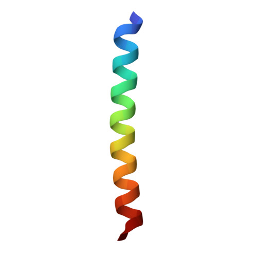
    While the application of cryogenic electron microscopy (cryo-EM) to helical polymers in biology has a long history, due to the huge number of helical macromolecular assemblies in viruses, bacteria, archaea, and eukaryotes, the use of cryo-EM to study synthetic soft matter noncovalent polymers has been much more limited. This has mainly been due to the lack of familiarity with cryo-EM in the materials science and chemistry communities, in contrast to the fact that cryo-EM was developed as a biological technique. Nevertheless, the relatively few structures of self-assembled peptide nanotubes and ribbons solved at near-atomic resolution by cryo-EM have demonstrated that cryo-EM should be the method of choice for a structural analysis of synthetic helical filaments. In addition, cryo-EM has also demonstrated that the self-assembly of soft matter polymers has enormous potential for polymorphism, something that may be obscured by techniques such as scattering and spectroscopy. These cryo-EM structures have revealed how far we currently are from being able to predict the structure of these polymers due to their chaotic self-assembly behavior.


  • Organizational Affiliation

    Department of Biochemistry and Molecular Genetics, University of Virginia, Charlottesville, Virginia 22908, United States.


Macromolecules
Find similar proteins by:  (by identity cutoff)  |  3D Structure
Entity ID: 1
MoleculeChains Sequence LengthOrganismDetailsImage
F1-N2 nanotube29synthetic constructMutation(s): 0 
Entity Groups  
Sequence Clusters30% Identity50% Identity70% Identity90% Identity95% Identity100% Identity
Sequence Annotations
Expand
  • Reference Sequence
Experimental Data & Validation

Experimental Data

  • Method: ELECTRON MICROSCOPY
  • Resolution: 3.40 Å
  • Aggregation State: FILAMENT 
  • Reconstruction Method: HELICAL 

Structure Validation

View Full Validation Report


Entry History & Funding Information

Deposition Data


Funding OrganizationLocationGrant Number
National Science Foundation (NSF, United States)United StatesNSF-DMR-1533958
National Institutes of Health/National Institute of General Medical Sciences (NIH/NIGMS)United StatesR35GM122510
National Institutes of Health/National Institute of General Medical Sciences (NIH/NIGMS)United StatesK99GM138756

Revision History  (Full details and data files)

  • Version 1.0: 2021-09-08
    Type: Initial release
  • Version 1.1: 2022-02-23
    Changes: Database references, Derived calculations
  • Version 1.2: 2022-03-02
    Changes: Database references
  • Version 1.3: 2022-09-28
    Changes: Database references